

ng Applications in Genetics
Detection of 9-bp
Deletion by SYBR
Green I-Based
Method
I'tJ
-
- .... u..
- 'tJ I
GI
IJ
C
GI
IJ
III
GI
:e..
o
~
2.62 .0u..
0,0Patient
Brother
2. bp del
,
I
I
I
I
I
I
I
I t '
54.0 ~.o
58.0
80.0
&2 .0
&4,0 oe.o ca.o 70.0 72 .0 74.0
Temperature (0C)
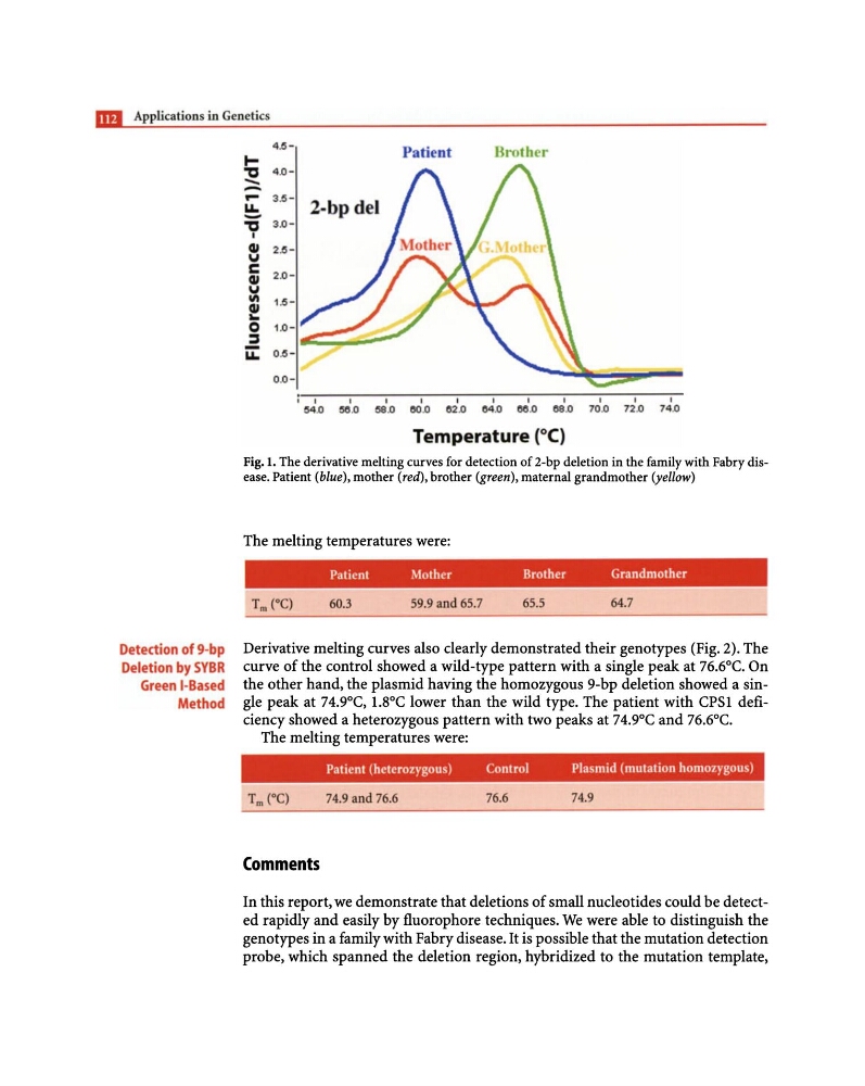
Fig. 1. The derivative melting curves for detection of 2-bp deletion in the family with Fabry disease. Patient (blue), mother (red), brother (green), maternal grandmother (yellow)
The melting temperatures were:
Patient
Mother
Brother
Grandmother
Tm (0C)
60.3
59.9 and 65,7
65.5
64.7
Derivative melting curves also clearly demonstrated their genotypes (Fig. 2). The
curve of the control showed a wild-type pattern with a single peak at 76.6°C. On
the other hand, the plasmid having the homozygous 9-bp deletion showed a single peak at 74.9°C, 1.8°C lower than the wild type. The patient with CPSl deficiency showed a heterozygous pattern with two peaks at 74.9°C and 76.6°C.
The melting temperatures were:
Patient (heterozygous)
Control
Plasmid (mutation homozygous)
74.9 and 76.6
76.6
74,9
Comments
In this report, we demonstrate that deletions of small nucleotides could be detected rapidly and easily by fluorophore techniques. We were able to distinguish the
genotypes in a family with Fabry disease. It is possible that the mutation detection
probe, which spanned the deletion region, hybridized to the mutation template,
Detection of 9-bp
Deletion by SYBR
Green I-Based
Method
I'tJ
-
- .... u..
- 'tJ I
GI
IJ
C
GI
IJ
III
GI
:e..
o
~
2.62 .0u..
0,0Patient
Brother
2. bp del
,
I
I
I
I
I
I
I
I t '
54.0 ~.o
58.0
80.0
&2 .0
&4,0 oe.o ca.o 70.0 72 .0 74.0
Temperature (0C)
Fig. 1. The derivative melting curves for detection of 2-bp deletion in the family with Fabry disease. Patient (blue), mother (red), brother (green), maternal grandmother (yellow)
The melting temperatures were:
Patient
Mother
Brother
Grandmother
Tm (0C)
60.3
59.9 and 65,7
65.5
64.7
Derivative melting curves also clearly demonstrated their genotypes (Fig. 2). The
curve of the control showed a wild-type pattern with a single peak at 76.6°C. On
the other hand, the plasmid having the homozygous 9-bp deletion showed a single peak at 74.9°C, 1.8°C lower than the wild type. The patient with CPSl deficiency showed a heterozygous pattern with two peaks at 74.9°C and 76.6°C.
The melting temperatures were:
Patient (heterozygous)
Control
Plasmid (mutation homozygous)
74.9 and 76.6
76.6
74,9
Comments
In this report, we demonstrate that deletions of small nucleotides could be detected rapidly and easily by fluorophore techniques. We were able to distinguish the
genotypes in a family with Fabry disease. It is possible that the mutation detection
probe, which spanned the deletion region, hybridized to the mutation template,