

Rapid GeDotyping of 2-bp and 9-bp Deletion Mutations Using the LightCycler Instrument IIIJ
10.0~
11.0"
8.0- - 9-bp del
-
u..
-
"
I
C»
'" C C»
'" III C» II. 0
:s
u..
8.05 .04 .03 .02 .00.0-,
I
71 .0
,
72.0
I
73.0
,
74.0
I
75.0
I
78.0
I
77.0
I
78.0
Temperature (O C)
I
711.0
I
80.0
I
81.0
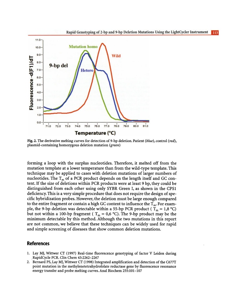
Fig. 2. The derivative melting curves for detection of 9-bp deletion. Patient (blue), control (red),
plasmid-containing homozygous deletion mutation (green)
forming a loop with the surplus nucleotides. Therefore, it melted off from the
mutation template at a lower temperature than from the wild-type template. This
technique may be applied to cases with deletion mutations of larger numbers of
nucleotides. The T m of a PCR product depends on the length itself and GC content. If the size of deletions within PCR products were at least 9 bp, they could be
distinguished from each other using only SYBR Green I, as shown in the CPS1
deficiency. This is a very simple procedure that does not require the design of specific hybridization probes. However, the deletion must be large enough compared
to the entire fragment or contain a high GC content to influence the T m' For example, the 9-bp deletion was detectable within a 55-bp PCR product ( Tm = 1,8 0c)
but not within a 100-bp fragment ( Tm = 0,6 °C). The 9-bp product may be the
minimum detectable by this method. Although the two mutations in this report
are not common, we believe that these techniques can be widely used for rapid
and simple screening of diseases that show common deletion mutations.
References
1. Lay MJ, Wittwer CT (1997) Real-time fluorescence genotyping of factor V Leiden during
. RapidCyde PCR. Clin Chern 43:2262-2267
2. Bernard PS, Lay MJ, Wittwer CT (1998) Integrated amplification and detection of the C677T
point mutation in the methylenetetrahydrofolate reductase gene by fluorescence resonance
energy transfer and probe melting curves. Anal Biochem 255:101-107
10.0~
11.0"
8.0- - 9-bp del
-
u..
-
"
I
C»
'" C C»
'" III C» II. 0
:s
u..
8.05 .04 .03 .02 .00.0-,
I
71 .0
,
72.0
I
73.0
,
74.0
I
75.0
I
78.0
I
77.0
I
78.0
Temperature (O C)
I
711.0
I
80.0
I
81.0
Fig. 2. The derivative melting curves for detection of 9-bp deletion. Patient (blue), control (red),
plasmid-containing homozygous deletion mutation (green)
forming a loop with the surplus nucleotides. Therefore, it melted off from the
mutation template at a lower temperature than from the wild-type template. This
technique may be applied to cases with deletion mutations of larger numbers of
nucleotides. The T m of a PCR product depends on the length itself and GC content. If the size of deletions within PCR products were at least 9 bp, they could be
distinguished from each other using only SYBR Green I, as shown in the CPS1
deficiency. This is a very simple procedure that does not require the design of specific hybridization probes. However, the deletion must be large enough compared
to the entire fragment or contain a high GC content to influence the T m' For example, the 9-bp deletion was detectable within a 55-bp PCR product ( Tm = 1,8 0c)
but not within a 100-bp fragment ( Tm = 0,6 °C). The 9-bp product may be the
minimum detectable by this method. Although the two mutations in this report
are not common, we believe that these techniques can be widely used for rapid
and simple screening of diseases that show common deletion mutations.
References
1. Lay MJ, Wittwer CT (1997) Real-time fluorescence genotyping of factor V Leiden during
. RapidCyde PCR. Clin Chern 43:2262-2267
2. Bernard PS, Lay MJ, Wittwer CT (1998) Integrated amplification and detection of the C677T
point mutation in the methylenetetrahydrofolate reductase gene by fluorescence resonance
energy transfer and probe melting curves. Anal Biochem 255:101-107