

8 Electronic Properties of Transition Metal-Benzene Sandwich Clusters
329
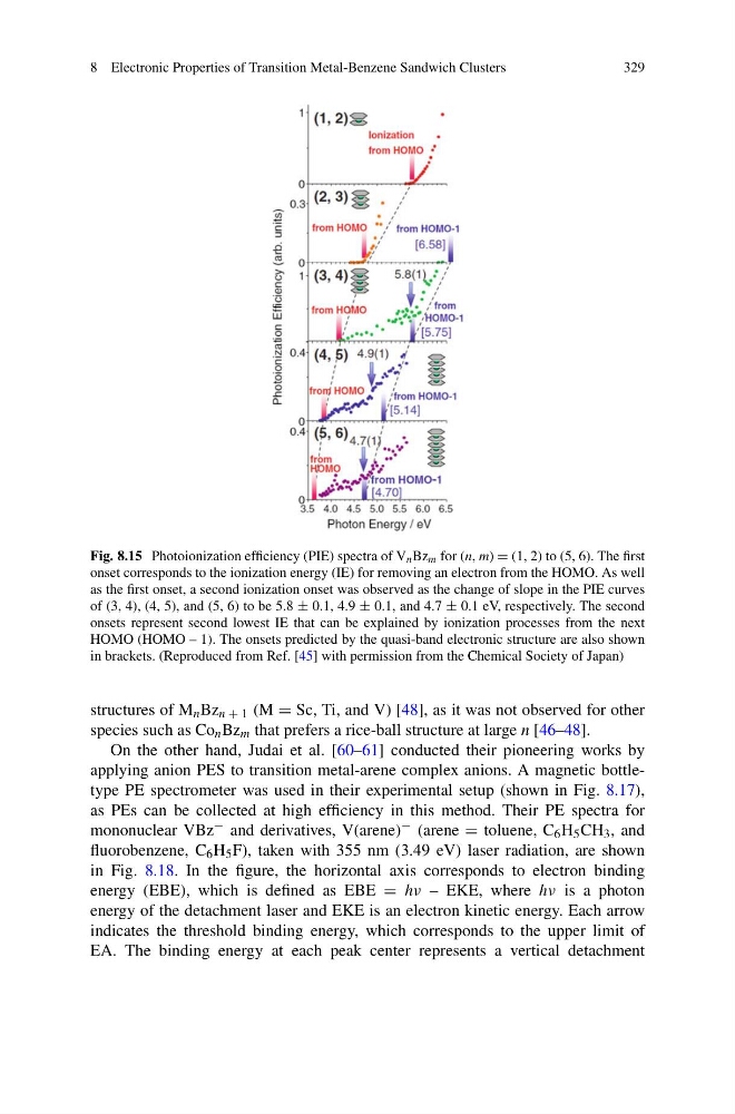
Fig. 8.15 Photoionization efficiency (PIE) spectra of V n Bz m for (n, m) = (1, 2) to (5, 6). The first
onset corresponds to the ionization energy (IE) for removing an electron from the HOMO. As well
as the first onset, a second ionization onset was observed as the change of slope in the PIE curves
of (3, 4), (4, 5), and (5, 6) to be 5.8 ± 0.1, 4.9 ± 0.1, and 4.7 ± 0.1 eV, respectively. The second
onsets represent second lowest IE that can be explained by ionization processes from the next
HOMO (HOMO – 1). The onsets predicted by the quasi-band electronic structure are also shown
in brackets. (Reproduced from Ref. [45] with permission from the Chemical Society of Japan)
structures of M n Bz n + 1 (M = Sc, Ti, and V) [48], as it was not observed for other
species such as Co n Bz m that prefers a rice-ball structure at large n [46–48].
On the other hand, Judai et al. [60–61] conducted their pioneering works by
applying anion PES to transition metal-arene complex anions. A magnetic bottletype PE spectrometer was used in their experimental setup (shown in Fig. 8.17),
as PEs can be collected at high efficiency in this method. Their PE spectra for
mononuclear VBz − and derivatives, V(arene) − (arene = toluene, C 6 H 5 CH 3 , and
fluorobenzene, C 6 H 5 F), taken with 355 nm (3.49 eV) laser radiation, are shown
in Fig. 8.18. In the figure, the horizontal axis corresponds to electron binding
energy (EBE), which is defined as EBE = hν – EKE, where hν is a photon
energy of the detachment laser and EKE is an electron kinetic energy. Each arrow
indicates the threshold binding energy, which corresponds to the upper limit of
EA. The binding energy at each peak center represents a vertical detachment
329
Fig. 8.15 Photoionization efficiency (PIE) spectra of V n Bz m for (n, m) = (1, 2) to (5, 6). The first
onset corresponds to the ionization energy (IE) for removing an electron from the HOMO. As well
as the first onset, a second ionization onset was observed as the change of slope in the PIE curves
of (3, 4), (4, 5), and (5, 6) to be 5.8 ± 0.1, 4.9 ± 0.1, and 4.7 ± 0.1 eV, respectively. The second
onsets represent second lowest IE that can be explained by ionization processes from the next
HOMO (HOMO – 1). The onsets predicted by the quasi-band electronic structure are also shown
in brackets. (Reproduced from Ref. [45] with permission from the Chemical Society of Japan)
structures of M n Bz n + 1 (M = Sc, Ti, and V) [48], as it was not observed for other
species such as Co n Bz m that prefers a rice-ball structure at large n [46–48].
On the other hand, Judai et al. [60–61] conducted their pioneering works by
applying anion PES to transition metal-arene complex anions. A magnetic bottletype PE spectrometer was used in their experimental setup (shown in Fig. 8.17),
as PEs can be collected at high efficiency in this method. Their PE spectra for
mononuclear VBz − and derivatives, V(arene) − (arene = toluene, C 6 H 5 CH 3 , and
fluorobenzene, C 6 H 5 F), taken with 355 nm (3.49 eV) laser radiation, are shown
in Fig. 8.18. In the figure, the horizontal axis corresponds to electron binding
energy (EBE), which is defined as EBE = hν – EKE, where hν is a photon
energy of the detachment laser and EKE is an electron kinetic energy. Each arrow
indicates the threshold binding energy, which corresponds to the upper limit of
EA. The binding energy at each peak center represents a vertical detachment