

110
S. Yanagisawa and I. Hamada
HOMO
LUMO
(a)
t 1
(b)
a′
c′
t 2
a′
c′
t 1
t 2
HOMO
LUMO
a′
b′
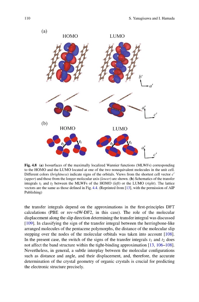
Fig. 4.8 (a) Isosurfaces of the maximally localized Wannier functions (MLWFs) corresponding
to the HOMO and the LUMO located at one of the two nonequivalent molecules in the unit cell.
Different colors (brightness) indicate signs of the orbitals. Views from the shortest cell vector c
(upper) and those from the longer molecular axis (lower) are shown. (b) Schematics of the transfer
integrals t 1 and t 2 between the MLWFs of the HOMO (left) or the LUMO (right). The lattice
vectors are the same as those defined in Fig. 4.4. (Reprinted from [13], with the permission of AIP
Publishing)
the transfer integrals depend on the approximations in the first-principles DFT
calculations (PBE or rev-vdW-DF2, in this case). The role of the molecular
displacement along the slip direction determining the transfer integral was discussed
[109]. In classifying the sign of the transfer integral between the herringbone-like
arranged molecules of the pentacene polymorphs, the distance of the molecular slip
stepping over the nodes of the molecular orbitals was taken into account [108].
In the present case, the switch of the signs of the transfer integrals t 1 and t 2 does
not affect the band structure within the tight-binding approximation [13, 106–108].
Nevertheless, in general, a subtle interplay between the molecular configurations
such as distance and angle, and their displacement, and, therefore, the accurate
determination of the crystal geometry of organic crystals is crucial for predicting
the electronic structure precisely.
S. Yanagisawa and I. Hamada
HOMO
LUMO
(a)
t 1
(b)
a′
c′
t 2
a′
c′
t 1
t 2
HOMO
LUMO
a′
b′
Fig. 4.8 (a) Isosurfaces of the maximally localized Wannier functions (MLWFs) corresponding
to the HOMO and the LUMO located at one of the two nonequivalent molecules in the unit cell.
Different colors (brightness) indicate signs of the orbitals. Views from the shortest cell vector c
(upper) and those from the longer molecular axis (lower) are shown. (b) Schematics of the transfer
integrals t 1 and t 2 between the MLWFs of the HOMO (left) or the LUMO (right). The lattice
vectors are the same as those defined in Fig. 4.4. (Reprinted from [13], with the permission of AIP
Publishing)
the transfer integrals depend on the approximations in the first-principles DFT
calculations (PBE or rev-vdW-DF2, in this case). The role of the molecular
displacement along the slip direction determining the transfer integral was discussed
[109]. In classifying the sign of the transfer integral between the herringbone-like
arranged molecules of the pentacene polymorphs, the distance of the molecular slip
stepping over the nodes of the molecular orbitals was taken into account [108].
In the present case, the switch of the signs of the transfer integrals t 1 and t 2 does
not affect the band structure within the tight-binding approximation [13, 106–108].
Nevertheless, in general, a subtle interplay between the molecular configurations
such as distance and angle, and their displacement, and, therefore, the accurate
determination of the crystal geometry of organic crystals is crucial for predicting
the electronic structure precisely.