

Biomarkers
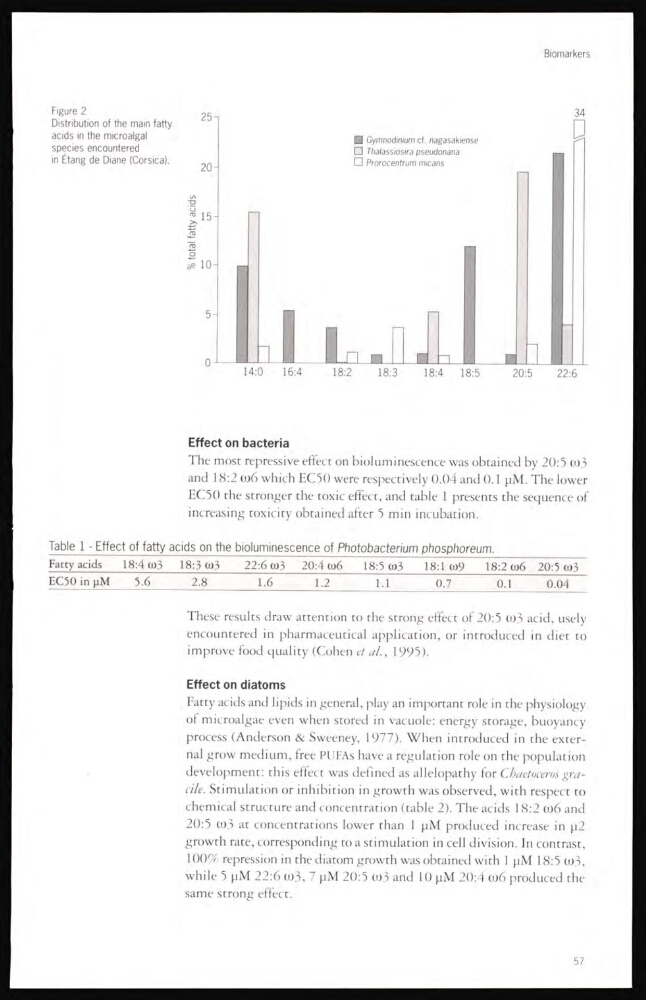
□ Gymnodinium cf. nagasakiense
□ Thalassiosira pseudonana
□ Prorocentrum micans
Figure 2
Distribution of the main fatty
acids in the microalgal
species encountered
in Étang de Diane (Corsica).
Effect on bacteria
The most repressive effect on bioluminescence was obtained by 20:5 ω3
and 18:2 ω6 which EC50 were respectively 0.04 and 0.1 pM. The lower
EC50 the stronger the toxic effect, and table 1 presents the sequence of
increasing toxicity obtained after 5 min incubation.
Table 1 - Effect of fatty acids on the bioluminescence of Photobacterium phosphoreum.
Fatty acids
18:4 ω3
18:3 ω3
22:6 ω3 20:4 ω6
18:5 ω3
18:1 ω9
18:2 ω6 20:5 ω3
EC50 in μM
5.6
2.8
1.6
1.2
1.1
0.7
0.1
0.04
These results draw attention to the strong effect of 20:5 0)3 acid, usely
encountered in pharmaceutical application, or introduced in diet to
improve food quality (Cohen et al., 1995).
Effect on diatoms
Fatty acids and lipids in general, play an important role in the physiology
of microalgae even when stored in vacuole: energy storage, buoyancy
process (Anderson & Sweeney, 1977). When introduced in the external grow medium, free PUFAs have a regulation role on the population
development: this effect was defined as allelopathy for Chaetoceros graale. Stimulation or inhibition in growth was observed, with respect to
chemical structure and concentration (table 2). The acids 18:2 ω6 and
20:5 0)3 at concentrations lower than 1 μM produced increase in μ2
growth rate, corresponding to a stimulation in cell division. In contrast,
100% repression in the diatom growth was obtained with 1 μM 18:5 ω3,
while 5 μM 22:6 ω3, 7 μM 20:5 ω3 and 10 μM 20: i ω6 produced the
same strong effect.
57
□ Gymnodinium cf. nagasakiense
□ Thalassiosira pseudonana
□ Prorocentrum micans
Figure 2
Distribution of the main fatty
acids in the microalgal
species encountered
in Étang de Diane (Corsica).
Effect on bacteria
The most repressive effect on bioluminescence was obtained by 20:5 ω3
and 18:2 ω6 which EC50 were respectively 0.04 and 0.1 pM. The lower
EC50 the stronger the toxic effect, and table 1 presents the sequence of
increasing toxicity obtained after 5 min incubation.
Table 1 - Effect of fatty acids on the bioluminescence of Photobacterium phosphoreum.
Fatty acids
18:4 ω3
18:3 ω3
22:6 ω3 20:4 ω6
18:5 ω3
18:1 ω9
18:2 ω6 20:5 ω3
EC50 in μM
5.6
2.8
1.6
1.2
1.1
0.7
0.1
0.04
These results draw attention to the strong effect of 20:5 0)3 acid, usely
encountered in pharmaceutical application, or introduced in diet to
improve food quality (Cohen et al., 1995).
Effect on diatoms
Fatty acids and lipids in general, play an important role in the physiology
of microalgae even when stored in vacuole: energy storage, buoyancy
process (Anderson & Sweeney, 1977). When introduced in the external grow medium, free PUFAs have a regulation role on the population
development: this effect was defined as allelopathy for Chaetoceros graale. Stimulation or inhibition in growth was observed, with respect to
chemical structure and concentration (table 2). The acids 18:2 ω6 and
20:5 0)3 at concentrations lower than 1 μM produced increase in μ2
growth rate, corresponding to a stimulation in cell division. In contrast,
100% repression in the diatom growth was obtained with 1 μM 18:5 ω3,
while 5 μM 22:6 ω3, 7 μM 20:5 ω3 and 10 μM 20: i ω6 produced the
same strong effect.
57