

Marine lipids
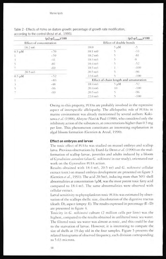
Table 2 - Effects of PUFAs on diatom growth: percentage of growth rate modification,
according to the control (Arzul et al., 1995).
(µ 2 -µ2 contr )/100
(µ2-µ2 contr )/100
Effect of concentration
Effect of double bonds
18:2 ω6
18:0
5 µM
-5
0.5 µM
+ 70
18:1 ω9
5
-33
1
+ 59
18:2 ω6
5
-81
3
-41
18:3 ω3
5
0
5
-81
18:4 ω3
5
-52
7
-69
18:5 ω3
1
-100
20:5 ω3
20:5 ω3
5
-96
0.5 µM
+ 52
22:6 ω5
5
-100
1
+ 83
Effect of chain length and unsaturation
3
-48
18:4 ω3
5 µM
-52
5
-96
20:4 ω6
10
-100
7
-99
20:5 0)3
5
-96
22:6 ω3
5
-100
Owing to this property, PUFAs are probably involved in the repressive
aspect of interspecific allelopathy. The allelopathic role of PUFAs in
marine environment was already mentionned by several authors: Kakisawa et al. (1988), Alstyne (Van) & Paul (1988), who considered only the
inhibitory action of the substances, at concentrations higher than 0.5 mg
per litre. This phenomenon constitutes an interesting explanation in
algal bloom formation (Gentien & Arzul, 1990).
Effect on embryos and larvae
The toxic effect of PUFAs was studied on mussel embryo and scallop
larva. Previous observations by Erard-Le Denn et al. (1990) on the malformation of scallop larvae, juveniles and adults induced by a bloom
of Gyrodinium aureolum (alias G. mikimotoi in our study), orientated our
work on the Gymnodium PUFA action.
Results obtained with 18:4 ω3, 20:5 ω3 and G. mikimotoi cellular
extract (extr.) on mussel embryo development are presented on figure 3
(Gentien et al., 1991 ). The acid 20:5ω3, inducing more than 50% shell
abnormalities at concentration lpM, was the most potent toxic fatty acid
compared to 18:4 ω3. The same abnormalities were observed with
cellular extract.
Larval sensitivity to phytoplankton toxic PUFAs was estimated by observation of the scallops shells: size, discoloration of the digestive tractus
(death: D), aspect (empty: E). The results expressed in percentage (E+D)
are presented in figure 4.
Toxicity in G. mikimotoi culture (2 million cells per litre) was the
highest, compared to the results obtained in unfiltered toxic sea water.
The filtered toxic sea water was almost as toxic, and this could be due
to the starvation of larvae. However, it is interesting to compare the
size of shells at 19 day old in the four samples. Figure 5 presents the
related histograms of observed frequency, each division corresponding
to 5.63 microns.
58
Table 2 - Effects of PUFAs on diatom growth: percentage of growth rate modification,
according to the control (Arzul et al., 1995).
(µ 2 -µ2 contr )/100
(µ2-µ2 contr )/100
Effect of concentration
Effect of double bonds
18:2 ω6
18:0
5 µM
-5
0.5 µM
+ 70
18:1 ω9
5
-33
1
+ 59
18:2 ω6
5
-81
3
-41
18:3 ω3
5
0
5
-81
18:4 ω3
5
-52
7
-69
18:5 ω3
1
-100
20:5 ω3
20:5 ω3
5
-96
0.5 µM
+ 52
22:6 ω5
5
-100
1
+ 83
Effect of chain length and unsaturation
3
-48
18:4 ω3
5 µM
-52
5
-96
20:4 ω6
10
-100
7
-99
20:5 0)3
5
-96
22:6 ω3
5
-100
Owing to this property, PUFAs are probably involved in the repressive
aspect of interspecific allelopathy. The allelopathic role of PUFAs in
marine environment was already mentionned by several authors: Kakisawa et al. (1988), Alstyne (Van) & Paul (1988), who considered only the
inhibitory action of the substances, at concentrations higher than 0.5 mg
per litre. This phenomenon constitutes an interesting explanation in
algal bloom formation (Gentien & Arzul, 1990).
Effect on embryos and larvae
The toxic effect of PUFAs was studied on mussel embryo and scallop
larva. Previous observations by Erard-Le Denn et al. (1990) on the malformation of scallop larvae, juveniles and adults induced by a bloom
of Gyrodinium aureolum (alias G. mikimotoi in our study), orientated our
work on the Gymnodium PUFA action.
Results obtained with 18:4 ω3, 20:5 ω3 and G. mikimotoi cellular
extract (extr.) on mussel embryo development are presented on figure 3
(Gentien et al., 1991 ). The acid 20:5ω3, inducing more than 50% shell
abnormalities at concentration lpM, was the most potent toxic fatty acid
compared to 18:4 ω3. The same abnormalities were observed with
cellular extract.
Larval sensitivity to phytoplankton toxic PUFAs was estimated by observation of the scallops shells: size, discoloration of the digestive tractus
(death: D), aspect (empty: E). The results expressed in percentage (E+D)
are presented in figure 4.
Toxicity in G. mikimotoi culture (2 million cells per litre) was the
highest, compared to the results obtained in unfiltered toxic sea water.
The filtered toxic sea water was almost as toxic, and this could be due
to the starvation of larvae. However, it is interesting to compare the
size of shells at 19 day old in the four samples. Figure 5 presents the
related histograms of observed frequency, each division corresponding
to 5.63 microns.
58