

Chemical Engineering and Processing - Process Intensification 163 (2021) 108359
8
glycone part cyclized between C-3 and C-6 of the aglycone; the presence
of a glucuronic acid on the oligosaccharide unit to the aglycone moiety
and the omnipresence of an aglycone of type Δ7,8-3β,6β-dihydroxysteroide Δ7,8-3β,6β-dihydroxysteroid (8-hydroxy-6-cholesta-8,14dien-23-one) [25].
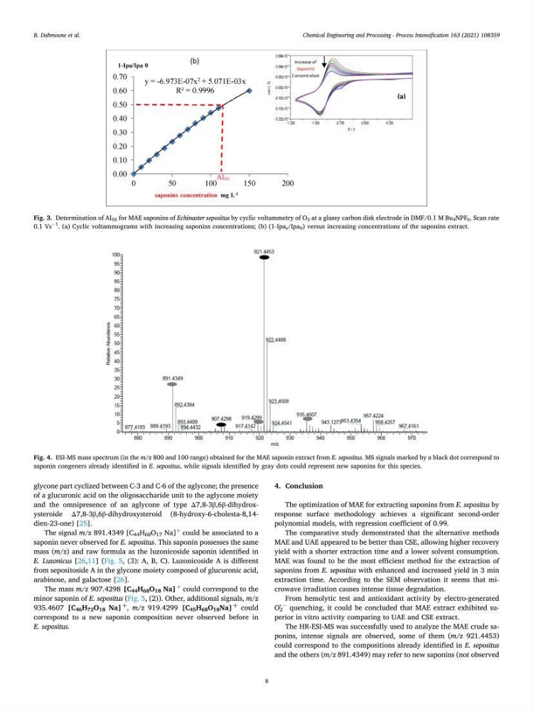
The signal m/z 891.4349 [C 44 H 68 O 17 Na]
+
could be associated to a
saponin never observed for E. sepositus. This saponin possesses the same
mass (m/z) and raw formula as the luzonicoside saponin identified in
E. Luzonicus [26,11] (Fig. 5, (3): A, B, C). Luzonicoside A is different
from sepositoside A in the glycone moiety composed of glucuronic acid,
arabinose, and galactose [26].
The mass m/z 907.4298 [C 44 H 68 O 18 Na]
+
could correspond to the
minor saponin of E. sepositus (Fig. 5, (2)). Other, additional signals, m/z
935.4607 [C 46 H 72 O 18 Na]
þ
, m/z 919.4299 [C 45 H 68 O 18 Na]
þ
could
correspond to a new saponin composition never observed before in
E. sepositus.
4. Conclusion
The optimization of MAE for extracting saponins from E. sepositus by
response surface methodology achieves a significant second-order
polynomial models, with regression coefficient of 0.99.
The comparative study demonstrated that the alternative methods
MAE and UAE appeared to be better than CSE, allowing higher recovery
yield with a shorter extraction time and a lower solvent consumption.
MAE was found to be the most efficient method for the extraction of
saponins from E. sepositus with enhanced and increased yield in 3 min
extraction time. According to the SEM observation it seems that microwave irradiation causes intense tissue degradation.
From hemolytic test and antioxidant activity by electro-generated
O 2
• −
quenching, it could be concluded that MAE extract exhibited superior in vitro activity comparing to UAE and CSE extract.
The HR-ESI-MS was successfully used to analyze the MAE crude saponins, intense signals are observed, some of them (m/z 921.4453)
could correspond to the compositions already identified in E. sepositus
and the others (m/z 891.4349) may refer to new saponins (not observed
Fig. 3. Determination of AI 50 for MAE saponins of Echinaster sepositus by cyclic voltammetry of O 2 at a glassy carbon disk electrode in DMF/0.1 M Bu 4 NPF 6 . Scan rate
0.1 Vs
− 1
. (a) Cyclic voltammograms with increasing saponins concentrations; (b) (1-Ipa s /Ipa 0 ) versus increasing concentrations of the saponins extract.
Fig. 4. ESI-MS mass spectrum (in the m/z 800 and 100 range) obtained for the MAE saponin extract from E. sepositus. MS signals marked by a black dot correspond to
saponin congeners already identified in E. sepositus, while signals identified by gray dots could represent new saponins for this species.
B. Dahmoune et al.
8
glycone part cyclized between C-3 and C-6 of the aglycone; the presence
of a glucuronic acid on the oligosaccharide unit to the aglycone moiety
and the omnipresence of an aglycone of type Δ7,8-3β,6β-dihydroxysteroide Δ7,8-3β,6β-dihydroxysteroid (8-hydroxy-6-cholesta-8,14dien-23-one) [25].
The signal m/z 891.4349 [C 44 H 68 O 17 Na]
+
could be associated to a
saponin never observed for E. sepositus. This saponin possesses the same
mass (m/z) and raw formula as the luzonicoside saponin identified in
E. Luzonicus [26,11] (Fig. 5, (3): A, B, C). Luzonicoside A is different
from sepositoside A in the glycone moiety composed of glucuronic acid,
arabinose, and galactose [26].
The mass m/z 907.4298 [C 44 H 68 O 18 Na]
+
could correspond to the
minor saponin of E. sepositus (Fig. 5, (2)). Other, additional signals, m/z
935.4607 [C 46 H 72 O 18 Na]
þ
, m/z 919.4299 [C 45 H 68 O 18 Na]
þ
could
correspond to a new saponin composition never observed before in
E. sepositus.
4. Conclusion
The optimization of MAE for extracting saponins from E. sepositus by
response surface methodology achieves a significant second-order
polynomial models, with regression coefficient of 0.99.
The comparative study demonstrated that the alternative methods
MAE and UAE appeared to be better than CSE, allowing higher recovery
yield with a shorter extraction time and a lower solvent consumption.
MAE was found to be the most efficient method for the extraction of
saponins from E. sepositus with enhanced and increased yield in 3 min
extraction time. According to the SEM observation it seems that microwave irradiation causes intense tissue degradation.
From hemolytic test and antioxidant activity by electro-generated
O 2
• −
quenching, it could be concluded that MAE extract exhibited superior in vitro activity comparing to UAE and CSE extract.
The HR-ESI-MS was successfully used to analyze the MAE crude saponins, intense signals are observed, some of them (m/z 921.4453)
could correspond to the compositions already identified in E. sepositus
and the others (m/z 891.4349) may refer to new saponins (not observed
Fig. 3. Determination of AI 50 for MAE saponins of Echinaster sepositus by cyclic voltammetry of O 2 at a glassy carbon disk electrode in DMF/0.1 M Bu 4 NPF 6 . Scan rate
0.1 Vs
− 1
. (a) Cyclic voltammograms with increasing saponins concentrations; (b) (1-Ipa s /Ipa 0 ) versus increasing concentrations of the saponins extract.
Fig. 4. ESI-MS mass spectrum (in the m/z 800 and 100 range) obtained for the MAE saponin extract from E. sepositus. MS signals marked by a black dot correspond to
saponin congeners already identified in E. sepositus, while signals identified by gray dots could represent new saponins for this species.
B. Dahmoune et al.