

376
particles are to the bulk signal. Flow cytometric analysis of individual cell properties has shed
some light on the matter.
1.2
A
1-0
cu
tl
CIS
1.0
tJ
1
en
...
.c:
0.8
~
't:I
S
!
.9
0.6
cu
(;r..
~ 0.4
§ !
cu
~
0.2
0.02
S
S
0
0.01
C')
...
::I
CIS
~ ~
B
cu 0.00
~
S
§
.9
cu -0.01
cu ~
III
! -0.02
12
18
24
30
36
42
48
Time (h)
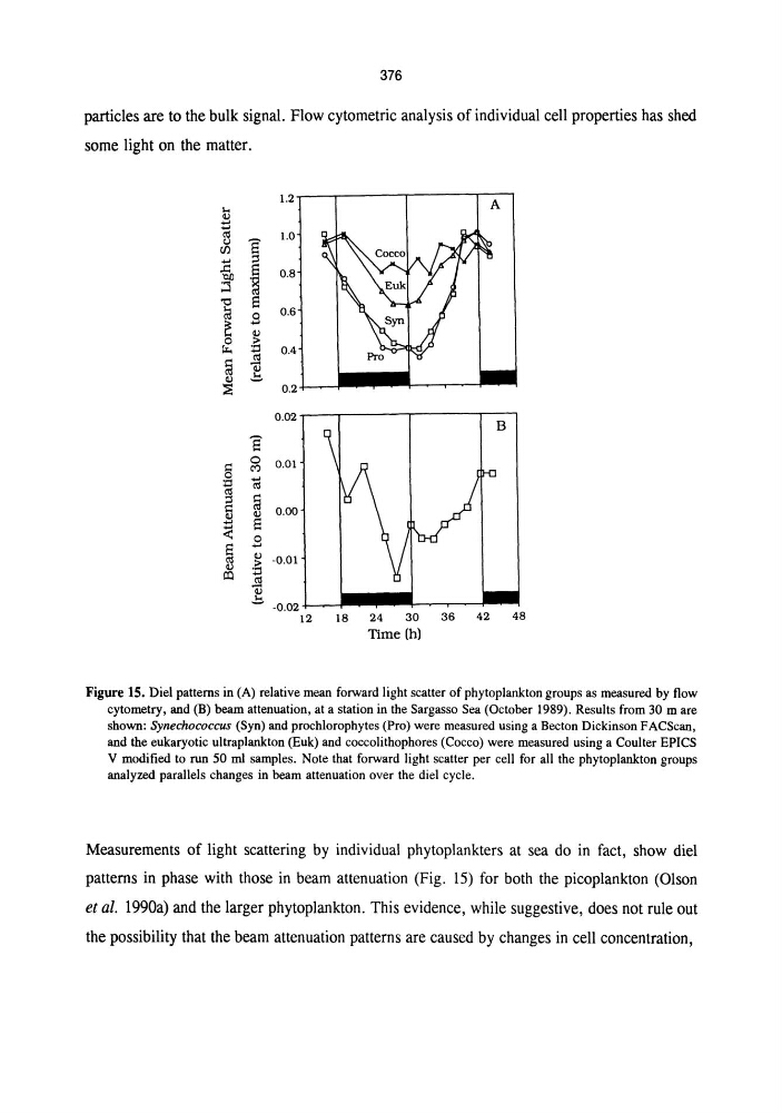
Figure IS. Diel patterns in (A) relative mean forward light scatter of phytoplankton groups as measured by flow
cytometry, and (B) beam attenuation, at a station in the Sargasso Sea (October 1989). Results from 30 mare
shown: Synechococcus (Syn) and prochlorophytes (Pro) were measured using a Becton Dickinson FACScan,
and the eukaryotic ultraplankton (Euk) and coccolithophores (Cocco) were measured using a Coulter EPICS
V modified to run 50 ml samples. Note that forward light scatter per cell for all the phytoplankton groups
analyzed parallels changes in beam attenuation over the diel cycle.
Measurements of light scattering by individual phytoplankters at sea do in fact, show diel
patterns in phase with those in beam attenuation (Fig. 15) for both the picoplankton (Olson
et al. 1990a) and the larger phytoplankton. This evidence, while suggestive, does not rule out
the possibility that the beam attenuation patterns are caused by changes in cell concentration,
particles are to the bulk signal. Flow cytometric analysis of individual cell properties has shed
some light on the matter.
1.2
A
1-0
cu
tl
CIS
1.0
tJ
1
en
...
.c:
0.8
~
't:I
S
!
.9
0.6
cu
(;r..
~ 0.4
§ !
cu
~
0.2
0.02
S
S
0
0.01
C')
...
::I
CIS
~ ~
B
cu 0.00
~
S
§
.9
cu -0.01
cu ~
III
! -0.02
12
18
24
30
36
42
48
Time (h)
Figure IS. Diel patterns in (A) relative mean forward light scatter of phytoplankton groups as measured by flow
cytometry, and (B) beam attenuation, at a station in the Sargasso Sea (October 1989). Results from 30 mare
shown: Synechococcus (Syn) and prochlorophytes (Pro) were measured using a Becton Dickinson FACScan,
and the eukaryotic ultraplankton (Euk) and coccolithophores (Cocco) were measured using a Coulter EPICS
V modified to run 50 ml samples. Note that forward light scatter per cell for all the phytoplankton groups
analyzed parallels changes in beam attenuation over the diel cycle.
Measurements of light scattering by individual phytoplankters at sea do in fact, show diel
patterns in phase with those in beam attenuation (Fig. 15) for both the picoplankton (Olson
et al. 1990a) and the larger phytoplankton. This evidence, while suggestive, does not rule out
the possibility that the beam attenuation patterns are caused by changes in cell concentration,