

377
5
~
A
« 4
U
tI.l
~
3
~
fi 2
f,l:l
~
«
~
0
30
TIME OF DAY (hours)
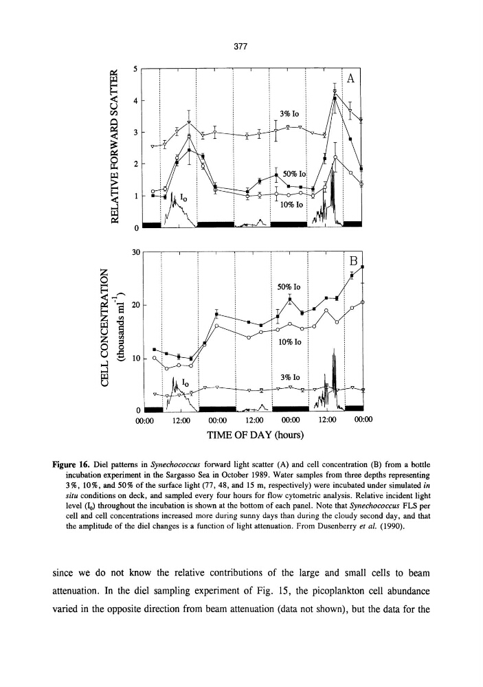
Figure 16. Diel patterns in Synechococcus fOIWard light scatter (A) and cell concentration (B) from a bottle
incubation experiment in the Sargasso Sea in October 1989. Water samples from three depths representing
3 %, 10%, and 50% of the surface light (77, 48, and 15 m, respectively) were incubated under simulated in
situ conditions on deck, and sampled every four hours for flow cytometric analysis. Relative incident light
level ~) throughout the incubation is shown at the bottom of each panel. Note that SYllechococcus FLS per
cell and cell concentrations increased more during sunny days than during the cloudy second day, and that
the amplitude of the diel changes is a function of light attenuation. From Dusenberry et al. (1990).
since we do not know the relative contributions of the large and small cells to beam
attenuation. In the diel sampling experiment of Fig. 15, the picoplankton cell abundance
varied in the opposite direction from beam attenuation (data not shown), but the data for the
5
~
A
« 4
U
tI.l
~
3
~
fi 2
f,l:l
~
«
~
0
30
TIME OF DAY (hours)
Figure 16. Diel patterns in Synechococcus fOIWard light scatter (A) and cell concentration (B) from a bottle
incubation experiment in the Sargasso Sea in October 1989. Water samples from three depths representing
3 %, 10%, and 50% of the surface light (77, 48, and 15 m, respectively) were incubated under simulated in
situ conditions on deck, and sampled every four hours for flow cytometric analysis. Relative incident light
level ~) throughout the incubation is shown at the bottom of each panel. Note that SYllechococcus FLS per
cell and cell concentrations increased more during sunny days than during the cloudy second day, and that
the amplitude of the diel changes is a function of light attenuation. From Dusenberry et al. (1990).
since we do not know the relative contributions of the large and small cells to beam
attenuation. In the diel sampling experiment of Fig. 15, the picoplankton cell abundance
varied in the opposite direction from beam attenuation (data not shown), but the data for the