

196
swelled human blood cell is viewed as a homogeneous sphere of cytoplasm surrounded by a
thin, homogeneous coating of membrane. The coated sphere model was used to calculate SII
between 1 degree and 15 degrees for Chinese hamster tissue culture cells (CHO), which are
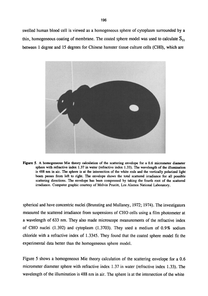
Figure S. A homogeneous Mie theory calculation of the scattering envelope for a 0.6 micrometer diameter
sphere with refractive index 1.37 in water (refractive index 1.33). The wavelength of the illumination
is 488 nm in air. The sphere is at the intersection of the white rods and the vertically polarized light
beam passes from left to right. The envelope shows the total scattered irradiance for all possible
scattering directions. The envelope has been compressed by taking the fourth root of the scattered
irradiance. Computer graphic courtesy of Melvin Prueitt, Los Alamos National Laboratory.
spherical and have concentric nuclei (Brunsting and Mullaney, 1972; 1974). The investigators
measured the scattered irradiance from suspensions of CHO cells using a film photometer at
a wavelength of 633 nm. They also made microscope measurements of the refractive index
of CRO nuclei (1.392) and cytoplasm (1.3703). They used a medium of 0.9% sodium
chloride with a refractive index of 1.3345. They found that the coated sphere model fit the
experimental data better than the homogeneous sphere model.
Figure 5 shows a homogeneous Mie theory calculation of the scattering envelope for a 0.6
micrometer diameter sphere with refractive index 1.37 in water (refractive index 1.33). The
wavelength of the illumination is 488 nm in air. The sphere is at the intersection of the white
swelled human blood cell is viewed as a homogeneous sphere of cytoplasm surrounded by a
thin, homogeneous coating of membrane. The coated sphere model was used to calculate SII
between 1 degree and 15 degrees for Chinese hamster tissue culture cells (CHO), which are
Figure S. A homogeneous Mie theory calculation of the scattering envelope for a 0.6 micrometer diameter
sphere with refractive index 1.37 in water (refractive index 1.33). The wavelength of the illumination
is 488 nm in air. The sphere is at the intersection of the white rods and the vertically polarized light
beam passes from left to right. The envelope shows the total scattered irradiance for all possible
scattering directions. The envelope has been compressed by taking the fourth root of the scattered
irradiance. Computer graphic courtesy of Melvin Prueitt, Los Alamos National Laboratory.
spherical and have concentric nuclei (Brunsting and Mullaney, 1972; 1974). The investigators
measured the scattered irradiance from suspensions of CHO cells using a film photometer at
a wavelength of 633 nm. They also made microscope measurements of the refractive index
of CRO nuclei (1.392) and cytoplasm (1.3703). They used a medium of 0.9% sodium
chloride with a refractive index of 1.3345. They found that the coated sphere model fit the
experimental data better than the homogeneous sphere model.
Figure 5 shows a homogeneous Mie theory calculation of the scattering envelope for a 0.6
micrometer diameter sphere with refractive index 1.37 in water (refractive index 1.33). The
wavelength of the illumination is 488 nm in air. The sphere is at the intersection of the white