

402
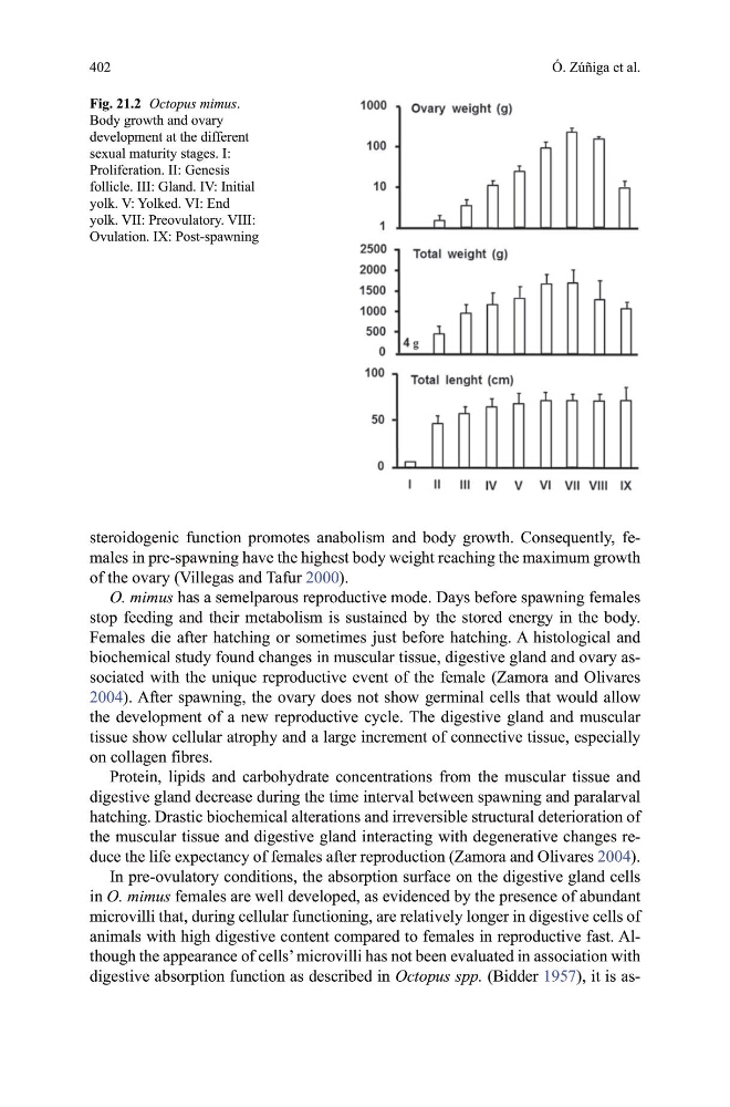
steroidogenic function promotes anabolism and body growth. Consequently, females in pre-spawning have the highest body weight reaching the maximum growth
of the ovary (Villegas and Tafur 2000).
O� mimus has a semelparous reproductive mode. Days before spawning females
stop feeding and their metabolism is sustained by the stored energy in the body.
Females die after hatching or sometimes just before hatching. A histological and
biochemical study found changes in muscular tissue, digestive gland and ovary associated with the unique reproductive event of the female (Zamora and Olivares
2004). After spawning, the ovary does not show germinal cells that would allow
the development of a new reproductive cycle. The digestive gland and muscular
tissue show cellular atrophy and a large increment of connective tissue, especially
on collagen fibres.
Protein, lipids and carbohydrate concentrations from the muscular tissue and
digestive gland decrease during the time interval between spawning and paralarval
hatching. Drastic biochemical alterations and irreversible structural deterioration of
the muscular tissue and digestive gland interacting with degenerative changes reduce the life expectancy of females after reproduction (Zamora and Olivares 2004).
In pre-ovulatory conditions, the absorption surface on the digestive gland cells
in O� mimus females are well developed, as evidenced by the presence of abundant
microvilli that, during cellular functioning, are relatively longer in digestive cells of
animals with high digestive content compared to females in reproductive fast. Although the appearance of cells’ microvilli has not been evaluated in association with
digestive absorption function as described in Octopus spp� (Bidder 1957), it is asFig. 21.2 Octopus mimus.
Body growth and ovary
development at the different
sexual maturity stages. I:
Proliferation. II: Genesis
follicle. III: Gland. IV: Initial
yolk. V: Yolked. VI: End
yolk. VII: Preovulatory. VIII:
Ovulation. IX: Post-spawning
Ó. Zúñiga et al.
steroidogenic function promotes anabolism and body growth. Consequently, females in pre-spawning have the highest body weight reaching the maximum growth
of the ovary (Villegas and Tafur 2000).
O� mimus has a semelparous reproductive mode. Days before spawning females
stop feeding and their metabolism is sustained by the stored energy in the body.
Females die after hatching or sometimes just before hatching. A histological and
biochemical study found changes in muscular tissue, digestive gland and ovary associated with the unique reproductive event of the female (Zamora and Olivares
2004). After spawning, the ovary does not show germinal cells that would allow
the development of a new reproductive cycle. The digestive gland and muscular
tissue show cellular atrophy and a large increment of connective tissue, especially
on collagen fibres.
Protein, lipids and carbohydrate concentrations from the muscular tissue and
digestive gland decrease during the time interval between spawning and paralarval
hatching. Drastic biochemical alterations and irreversible structural deterioration of
the muscular tissue and digestive gland interacting with degenerative changes reduce the life expectancy of females after reproduction (Zamora and Olivares 2004).
In pre-ovulatory conditions, the absorption surface on the digestive gland cells
in O� mimus females are well developed, as evidenced by the presence of abundant
microvilli that, during cellular functioning, are relatively longer in digestive cells of
animals with high digestive content compared to females in reproductive fast. Although the appearance of cells’ microvilli has not been evaluated in association with
digestive absorption function as described in Octopus spp� (Bidder 1957), it is asFig. 21.2 Octopus mimus.
Body growth and ovary
development at the different
sexual maturity stages. I:
Proliferation. II: Genesis
follicle. III: Gland. IV: Initial
yolk. V: Yolked. VI: End
yolk. VII: Preovulatory. VIII:
Ovulation. IX: Post-spawning
Ó. Zúñiga et al.