

21 Octopus mimus
401
females present in refuges from the spawning to the end of the parental care of the
embryos. Females weighing more than 890 g are found on different stages of sexual
maturity, which is determinate by the accumulation of yolk, follicle development
and ovulation (Olivares et al. 1996).
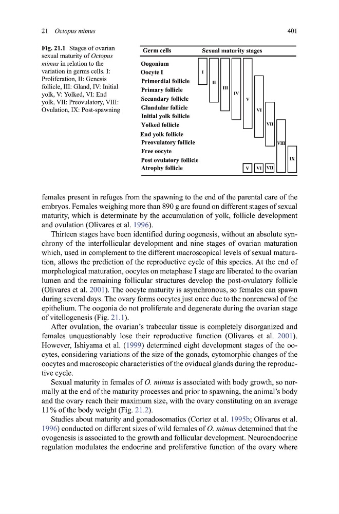
Thirteen stages have been identified during oogenesis, without an absolute synchrony of the interfollicular development and nine stages of ovarian maturation
which, used in complement to the different macroscopical levels of sexual maturation, allows the prediction of the reproductive cycle of this species. At the end of
morphological maturation, oocytes on metaphase I stage are liberated to the ovarian
lumen and the remaining follicular structures develop the post-ovulatory follicle
(Olivares et al. 2001). The oocyte maturity is asynchronous, so females can spawn
during several days. The ovary forms oocytes just once due to the nonrenewal of the
epithelium. The oogonia do not proliferate and degenerate during the ovarian stage
of vitellogenesis (Fig. 21.1).
After ovulation, the ovarian’s trabecular tissue is completely disorganized and
females unquestionably lose their reproductive function (Olivares et al. 2001).
However, Ishiyama et al. (1999) determined eight development stages of the oocytes, considering variations of the size of the gonads, cytomorphic changes of the
oocytes and macroscopic characteristics of the oviducal glands during the reproductive cycle.
Sexual maturity in females of O� mimus is associated with body growth, so normally at the end of the maturity processes and prior to spawning, the animal’s body
and the ovary reach their maximum size, with the ovary constituting on an average
11 % of the body weight (Fig. 21.2).
Studies about maturity and gonadosomatics (Cortez et al. 1995b; Olivares et al.
1996) conducted on different sizes of wild females of O� mimus determined that the
ovogenesis is associated to the growth and follicular development. Neuroendocrine
regulation modulates the endocrine and proliferative function of the ovary where
*HUP FHOOV
6H[XDOPDWXULW\ VWDJHV
9,,,
,;
9,,
9,
9
,9
,,,
,,
,
9
9, 9,,
2RJRQLXP
2RF\WH ,
3ULPRUGLDOIROOLFOH
3ULPDU\ IROOLFOH
6HFXQGDU\ IROOLFOH
*ODQGXODUIROOLFOH
,QLWLDO \RON IROOLFOH
)UHHRRF\WH
3RVWRYXODWRU\ IROOLFOH
$WURSK\ IROOLFOH
(QG \RON IROOLFOH
Fig. 21.1 Stages of ovarian
sexual maturity of Octopus
mimus in relation to the
variation in germs cells. I:
Proliferation, II: Genesis
follicle, III: Gland, IV: Initial
yolk, V: Yolked, VI: End
yolk, VII: Preovulatory, VIII:
Ovulation, IX: Post-spawning
401
females present in refuges from the spawning to the end of the parental care of the
embryos. Females weighing more than 890 g are found on different stages of sexual
maturity, which is determinate by the accumulation of yolk, follicle development
and ovulation (Olivares et al. 1996).
Thirteen stages have been identified during oogenesis, without an absolute synchrony of the interfollicular development and nine stages of ovarian maturation
which, used in complement to the different macroscopical levels of sexual maturation, allows the prediction of the reproductive cycle of this species. At the end of
morphological maturation, oocytes on metaphase I stage are liberated to the ovarian
lumen and the remaining follicular structures develop the post-ovulatory follicle
(Olivares et al. 2001). The oocyte maturity is asynchronous, so females can spawn
during several days. The ovary forms oocytes just once due to the nonrenewal of the
epithelium. The oogonia do not proliferate and degenerate during the ovarian stage
of vitellogenesis (Fig. 21.1).
After ovulation, the ovarian’s trabecular tissue is completely disorganized and
females unquestionably lose their reproductive function (Olivares et al. 2001).
However, Ishiyama et al. (1999) determined eight development stages of the oocytes, considering variations of the size of the gonads, cytomorphic changes of the
oocytes and macroscopic characteristics of the oviducal glands during the reproductive cycle.
Sexual maturity in females of O� mimus is associated with body growth, so normally at the end of the maturity processes and prior to spawning, the animal’s body
and the ovary reach their maximum size, with the ovary constituting on an average
11 % of the body weight (Fig. 21.2).
Studies about maturity and gonadosomatics (Cortez et al. 1995b; Olivares et al.
1996) conducted on different sizes of wild females of O� mimus determined that the
ovogenesis is associated to the growth and follicular development. Neuroendocrine
regulation modulates the endocrine and proliferative function of the ovary where
*HUP FHOOV
6H[XDOPDWXULW\ VWDJHV
9,,,
,;
9,,
9,
9
,9
,,,
,,
,
9
9, 9,,
2RJRQLXP
2RF\WH ,
3ULPRUGLDOIROOLFOH
3ULPDU\ IROOLFOH
6HFXQGDU\ IROOLFOH
*ODQGXODUIROOLFOH
,QLWLDO \RON IROOLFOH
)UHHRRF\WH
3RVWRYXODWRU\ IROOLFOH
$WURSK\ IROOLFOH
(QG \RON IROOLFOH
Fig. 21.1 Stages of ovarian
sexual maturity of Octopus
mimus in relation to the
variation in germs cells. I:
Proliferation, II: Genesis
follicle, III: Gland, IV: Initial
yolk, V: Yolked, VI: End
yolk, VII: Preovulatory, VIII:
Ovulation, IX: Post-spawning