

Hormones involved in acoustic communication include steroids and peptides. Steroid hormones, such as androgens and estrogens released from
the gonads, are small lipophilic molecules that readily diffuse through cell
membranes. In contrast, peptide hormones, such as AVP released from
hypothalamic neurons, are hydrophilic molecules that cannot readily pass
through cell membranes. Consequently, receptors for these two types of
hormones are localized differently: steroid receptors are found in the cytoplasm and/or nucleus of a cell, whereas peptide receptors are in the cell
membrane with an extracellular hormone-binding domain.
2.2.1. Steroid Hormones
The primary action of steroid hormones is to modify gene expression in
target cells. Steroid receptors are proteins localized in the cytoplasm/
278
A. Yamaguchi and D.B. Kelley
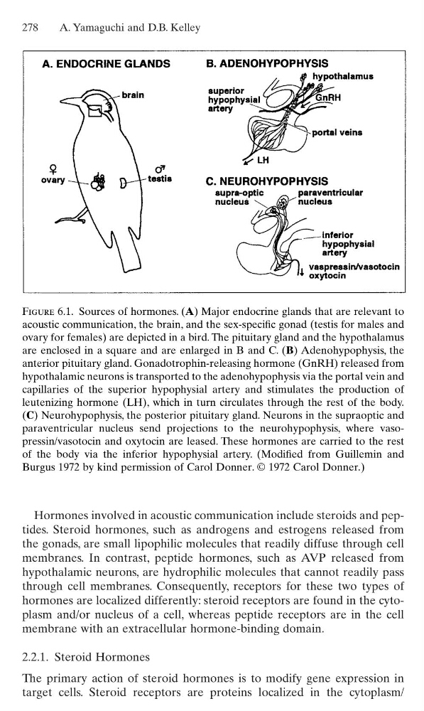
Figure 6.1. Sources of hormones. (A) Major endocrine glands that are relevant to
acoustic communication, the brain, and the sex-specific gonad (testis for males and
ovary for females) are depicted in a bird. The pituitary gland and the hypothalamus
are enclosed in a square and are enlarged in B and C. (B) Adenohypophysis, the
anterior pituitary gland. Gonadotrophin-releasing hormone (GnRH) released from
hypothalamic neurons is transported to the adenohypophysis via the portal vein and
capillaries of the superior hypophysial artery and stimulates the production of
leutenizing hormone (LH), which in turn circulates through the rest of the body.
(C) Neurohypophysis, the posterior pituitary gland. Neurons in the supraoptic and
paraventricular nucleus send projections to the neurohypophysis, where vasopressin/vasotocin and oxytocin are leased. These hormones are carried to the rest
of the body via the inferior hypophysial artery. (Modified from Guillemin and
Burgus 1972 by kind permission of Carol Donner. © 1972 Carol Donner.)
the gonads, are small lipophilic molecules that readily diffuse through cell
membranes. In contrast, peptide hormones, such as AVP released from
hypothalamic neurons, are hydrophilic molecules that cannot readily pass
through cell membranes. Consequently, receptors for these two types of
hormones are localized differently: steroid receptors are found in the cytoplasm and/or nucleus of a cell, whereas peptide receptors are in the cell
membrane with an extracellular hormone-binding domain.
2.2.1. Steroid Hormones
The primary action of steroid hormones is to modify gene expression in
target cells. Steroid receptors are proteins localized in the cytoplasm/
278
A. Yamaguchi and D.B. Kelley
Figure 6.1. Sources of hormones. (A) Major endocrine glands that are relevant to
acoustic communication, the brain, and the sex-specific gonad (testis for males and
ovary for females) are depicted in a bird. The pituitary gland and the hypothalamus
are enclosed in a square and are enlarged in B and C. (B) Adenohypophysis, the
anterior pituitary gland. Gonadotrophin-releasing hormone (GnRH) released from
hypothalamic neurons is transported to the adenohypophysis via the portal vein and
capillaries of the superior hypophysial artery and stimulates the production of
leutenizing hormone (LH), which in turn circulates through the rest of the body.
(C) Neurohypophysis, the posterior pituitary gland. Neurons in the supraoptic and
paraventricular nucleus send projections to the neurohypophysis, where vasopressin/vasotocin and oxytocin are leased. These hormones are carried to the rest
of the body via the inferior hypophysial artery. (Modified from Guillemin and
Burgus 1972 by kind permission of Carol Donner. © 1972 Carol Donner.)