

344
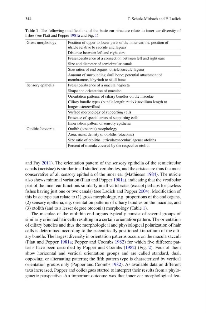
Table 1 The following modifi cations of the basic ear structure relate to inner ear diversity of
fi shes (see Platt and Popper 1981a and Fig. 1 )
Gross morphology
Position of upper to lower parts of the inner ear, i.e. position of
utricle relative to saccule and lagena
Distance between left and right ears
Presence/absence of a connection between left and right ears
Size and diameter of semicircular canals
Size ratios of end organs: utricle:saccule:lagena
Amount of surrounding skull bone; potential attachment of
membranous labyrinth to skull bone
Sensory epithelia
Presence/absence of a macula neglecta
Shape and orientation of maculae
Orientation patterns of ciliary bundles on the maculae
Ciliary bundle types (bundle length; ratio kinocilium length to
longest stereovillus)
Surface morphology of supporting cells
Presence of special areas of supporting cells
Innervation pattern of sensory epithelia
Otoliths/otoconia
Otolith (otoconia) morphology
Area, mass, density of otoliths (otoconia)
Size ratio of otoliths: utricular:saccular:lagenar otoliths
Percent of macula covered by the respective otolith
and Fay 2011 ). The orientation pattern of the sensory epithelia of the semicircular
canals (=cristae) is similar in all studied vertebrates, and the cristae are thus the most
conservative of all sensory epithelia of the inner ear (Mathiesen 1984 ). The utricle
also shows minimal variation (Platt and Popper 1981a ), indicating that the vestibular
part of the inner ear functions similarly in all vertebrates (except perhaps for jawless
fi shes having just one or two canals) (see Ladich and Popper 2004 ). Modifi cation of
this basic type can relate to (1) gross morphology, e.g. proportions of the end organs,
(2) sensory epithelia, e.g. orientation patterns of ciliary bundles on the maculae, and
(3) otolith (and to a lesser degree otoconia) morphology (Table 1 ).
The maculae of the otolithic end organs typically consist of several groups of
similarly oriented hair cells resulting in a certain orientation pattern. The orientation
of ciliary bundles and thus the morphological and physiological polarization of hair
cells is determined according to the eccentrically positioned kinocilium of the ciliary bundle. The largest diversity in orientation patterns occurs on the macula sacculi
(Platt and Popper 1981a ; Popper and Coombs 1982 ) for which fi ve different patterns have been described by Popper and Coombs ( 1982 ) (Fig. 2 ). Four of them
show horizontal and vertical orientation groups and are called standard, dual,
opposing, or alternating patterns; the fi fth pattern type is characterized by vertical
orientation groups only (Popper and Coombs 1982 ). As available data on different
taxa increased, Popper and colleagues started to interpret their results from a phylogenetic perspective. An important outcome was that inner ear morphological feaT. Schulz-Mirbach and F. Ladich
Table 1 The following modifi cations of the basic ear structure relate to inner ear diversity of
fi shes (see Platt and Popper 1981a and Fig. 1 )
Gross morphology
Position of upper to lower parts of the inner ear, i.e. position of
utricle relative to saccule and lagena
Distance between left and right ears
Presence/absence of a connection between left and right ears
Size and diameter of semicircular canals
Size ratios of end organs: utricle:saccule:lagena
Amount of surrounding skull bone; potential attachment of
membranous labyrinth to skull bone
Sensory epithelia
Presence/absence of a macula neglecta
Shape and orientation of maculae
Orientation patterns of ciliary bundles on the maculae
Ciliary bundle types (bundle length; ratio kinocilium length to
longest stereovillus)
Surface morphology of supporting cells
Presence of special areas of supporting cells
Innervation pattern of sensory epithelia
Otoliths/otoconia
Otolith (otoconia) morphology
Area, mass, density of otoliths (otoconia)
Size ratio of otoliths: utricular:saccular:lagenar otoliths
Percent of macula covered by the respective otolith
and Fay 2011 ). The orientation pattern of the sensory epithelia of the semicircular
canals (=cristae) is similar in all studied vertebrates, and the cristae are thus the most
conservative of all sensory epithelia of the inner ear (Mathiesen 1984 ). The utricle
also shows minimal variation (Platt and Popper 1981a ), indicating that the vestibular
part of the inner ear functions similarly in all vertebrates (except perhaps for jawless
fi shes having just one or two canals) (see Ladich and Popper 2004 ). Modifi cation of
this basic type can relate to (1) gross morphology, e.g. proportions of the end organs,
(2) sensory epithelia, e.g. orientation patterns of ciliary bundles on the maculae, and
(3) otolith (and to a lesser degree otoconia) morphology (Table 1 ).
The maculae of the otolithic end organs typically consist of several groups of
similarly oriented hair cells resulting in a certain orientation pattern. The orientation
of ciliary bundles and thus the morphological and physiological polarization of hair
cells is determined according to the eccentrically positioned kinocilium of the ciliary bundle. The largest diversity in orientation patterns occurs on the macula sacculi
(Platt and Popper 1981a ; Popper and Coombs 1982 ) for which fi ve different patterns have been described by Popper and Coombs ( 1982 ) (Fig. 2 ). Four of them
show horizontal and vertical orientation groups and are called standard, dual,
opposing, or alternating patterns; the fi fth pattern type is characterized by vertical
orientation groups only (Popper and Coombs 1982 ). As available data on different
taxa increased, Popper and colleagues started to interpret their results from a phylogenetic perspective. An important outcome was that inner ear morphological feaT. Schulz-Mirbach and F. Ladich