

Aeromonas salmonicida subspecies salmonicida is a
Gram negative, non-motile, facultative anaerobic rod. Pathogenicity is dependent on the so-called A-layer, an external
surface layer mainly composed of the A-protein that
provides protection against the defence mechanisms of the
host. At least 25 extracellular products are released during
growth and responsible for the tissue damage-degeneration
and ultimately, the death of the fish. Primary isolation of the
pathogen can be achieved from the kidney and other organs
on TSA or brain heart infusion agar (BHIA) at 22
C. Most
strains are oxidase positive and produce a water-soluble
brown pigment on medium containing tryptone. A less common and atypical strain of this pathogen is the subspecies
achromogenes, which does not produce pigment under standard incubation conditions and requires additional biochemical testing to differentiate. Diagnosis of A. salmonicida is
based upon gross and histopathological lesions, isolation of
the causative agent or its identification through immunohistochemistry, serology or molecular tools. In haematoxylin
and eosin (H&E) sections the appearance of the bacteria is
almost pathognomonic (see Fig. 6.5).
6.3
Listonella anguillarum
Listonella (Vibrio) anguillarum causes a haemorrhagic
septicaemia, an economical important disease affecting
salmonids in salt and brackish waters, particularly in the summer months at temperatures above 10
C. However, infections
with Vibrio ordalii and other Vibrio spp. are often collectively
termed ‘vibriosis’, and may cause disease with similar clinical
and pathological manifestation as L. anguillarum. Clinical
signs and pathological lesions may be variable, but similar to
several other Gram negative septicaemias, and dependant on
water temperature, fish age and pathogen virulence. L.
anguillarum may be present among the normal gut microflora
of healthy fish and clinical outbreaks triggered only by stress
activated virulent strains present in the gastrointestinal tract.
Poor water quality and rapid temperature changes can also
activate infection which then spreads horizontally. External
signs of disease include dark skin coloration, anorexia, pale
gills with increased mucous, periorbital oedema, swollen vent
(Fig. 6.7) and haemorrhage near the base of the pectoral and
pelvic fins. V. ordalli shows a predilection for muscle and skin
with resulting haemorrhage. Extensive multifocal liquefactive
muscle necrosis and haemorrhage with large numbers of bacteria is common (Fig. 6.8). Dermal or subdermal skin lesions
are often coupled to hyperaemia and haemorrhage, and may be
linked to occasional haemorrhagic ‘boil’ lesions in the muscle.
These may rupture and release blood and bacteria to the
surrounding water. Internally, petechiae may be present on
the peritoneal surface and in internal organs. The liver is
generally swollen and some fish show petechiae (Fig. 6.9).
Colonies of the bacterium are also found throughout the
Fig. 6.3 Furunculosis in farmed rainbow trout broodstock; subcutaneous boil lesions
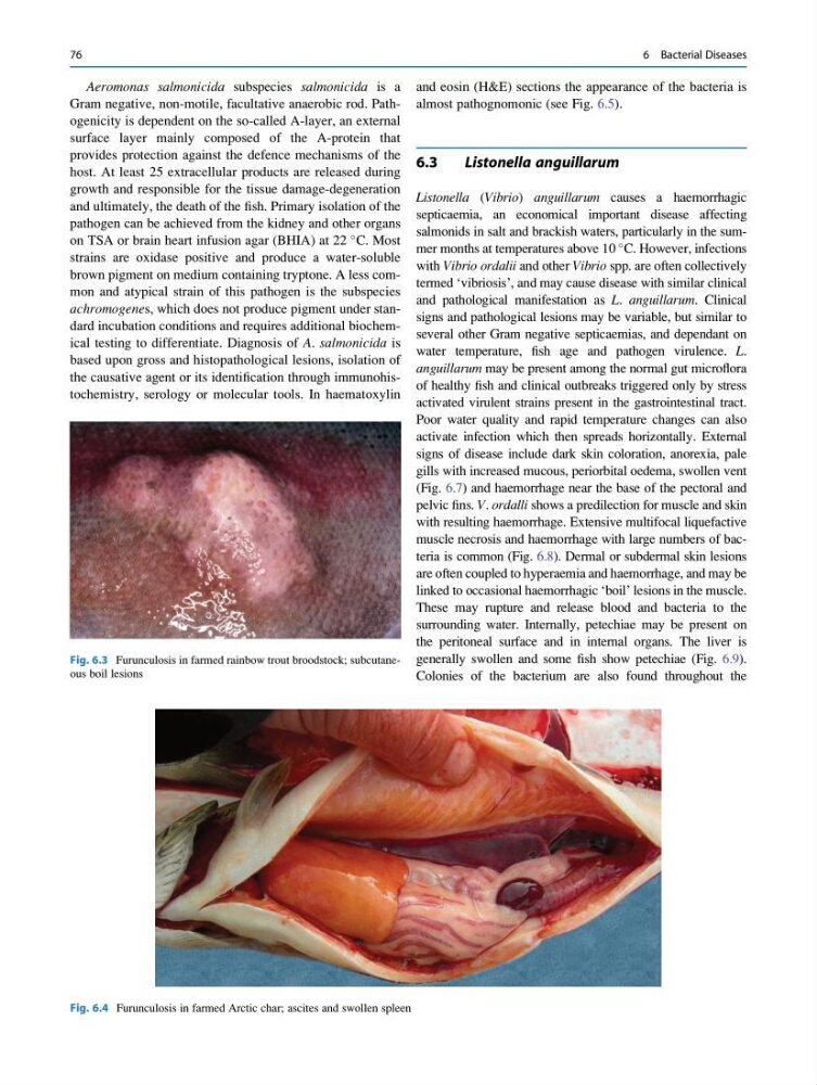
Fig. 6.4 Furunculosis in farmed Arctic char; ascites and swollen spleen
76
6 Bacterial Diseases
Gram negative, non-motile, facultative anaerobic rod. Pathogenicity is dependent on the so-called A-layer, an external
surface layer mainly composed of the A-protein that
provides protection against the defence mechanisms of the
host. At least 25 extracellular products are released during
growth and responsible for the tissue damage-degeneration
and ultimately, the death of the fish. Primary isolation of the
pathogen can be achieved from the kidney and other organs
on TSA or brain heart infusion agar (BHIA) at 22
C. Most
strains are oxidase positive and produce a water-soluble
brown pigment on medium containing tryptone. A less common and atypical strain of this pathogen is the subspecies
achromogenes, which does not produce pigment under standard incubation conditions and requires additional biochemical testing to differentiate. Diagnosis of A. salmonicida is
based upon gross and histopathological lesions, isolation of
the causative agent or its identification through immunohistochemistry, serology or molecular tools. In haematoxylin
and eosin (H&E) sections the appearance of the bacteria is
almost pathognomonic (see Fig. 6.5).
6.3
Listonella anguillarum
Listonella (Vibrio) anguillarum causes a haemorrhagic
septicaemia, an economical important disease affecting
salmonids in salt and brackish waters, particularly in the summer months at temperatures above 10
C. However, infections
with Vibrio ordalii and other Vibrio spp. are often collectively
termed ‘vibriosis’, and may cause disease with similar clinical
and pathological manifestation as L. anguillarum. Clinical
signs and pathological lesions may be variable, but similar to
several other Gram negative septicaemias, and dependant on
water temperature, fish age and pathogen virulence. L.
anguillarum may be present among the normal gut microflora
of healthy fish and clinical outbreaks triggered only by stress
activated virulent strains present in the gastrointestinal tract.
Poor water quality and rapid temperature changes can also
activate infection which then spreads horizontally. External
signs of disease include dark skin coloration, anorexia, pale
gills with increased mucous, periorbital oedema, swollen vent
(Fig. 6.7) and haemorrhage near the base of the pectoral and
pelvic fins. V. ordalli shows a predilection for muscle and skin
with resulting haemorrhage. Extensive multifocal liquefactive
muscle necrosis and haemorrhage with large numbers of bacteria is common (Fig. 6.8). Dermal or subdermal skin lesions
are often coupled to hyperaemia and haemorrhage, and may be
linked to occasional haemorrhagic ‘boil’ lesions in the muscle.
These may rupture and release blood and bacteria to the
surrounding water. Internally, petechiae may be present on
the peritoneal surface and in internal organs. The liver is
generally swollen and some fish show petechiae (Fig. 6.9).
Colonies of the bacterium are also found throughout the
Fig. 6.3 Furunculosis in farmed rainbow trout broodstock; subcutaneous boil lesions
Fig. 6.4 Furunculosis in farmed Arctic char; ascites and swollen spleen
76
6 Bacterial Diseases