

5.8
Hirame Rhabdovirus
Hirame rhabdovirus (HRV) was first reported from moribund Japanese flounder in 1984, and subsequently the virus
has been reported as pathogenic to species of bream. The
first description of HRV in Europe occurred in grayling and
brown trout in a farm in Poland, raising concerns that spread
may have occurred from Asiatic countries and there was
potential for emergence in freshwater fish.
Experimental studies involving salmonid fish (i.e. rainbow trout, chum, coho, masu and pink salmon) have been
carried out. The highest virus titre was obtained from
rainbow trout infected following direct transmission through
the water. Histologically, the experimentally HRV infected
rainbow trout showed necrosis and haemorrhage in the kidney and spleen and the skeletal muscle revealed hyperaemia
and haemorrhage. Due to similarities with VHS, this
becomes an important differential diagnosis for farmed rainbow trout. A qRT-PCR assay represents a reliable, specific
and sensitive tool for the quantitative diagnosis of HRV in
fish samples. Further work is required to assess if the virus is
spreading and its potential impact on farmed fish.
5.9
Salmonid Alphavirus (Pancreas
and Sleeping Disease)
Salmonid alphavirus is responsible for a serious infectious
disease affecting farmed Atlantic salmon (Pancreas disease,
PD), and rainbow trout (Sleeping disease, SD). It was first
reported in Scotland in 1976 in salmon followed by
observations in Norway and Ireland. The rainbow trout
condition was recognised by 1994 in France as a similar
disease and the confirmation of a viral aetiologies came
soon after with the isolation of the first alphavirus from
fish (PD) in Ireland (1995) and of SD, in France (1997).
Evidence shows that SAV represents an atypical and new
member of the genus Alphavirus within the family
Togaviridae. Currently six subtypes have been described,
and all of them affects sea water salmon (SAV 1, 2, 3, 4, 5
and 6), however only SAV 3 and recently SAV 2 have been
recorded in farmed salmon in Norway. The rainbow trout
fresh water virus is categorised as SAV-2, and SAV-2 marine
strains have been reported from salmon in Scotland. SAV
infections are recognised as a major disease problem in
farmed salmonids in the UK, Ireland and Norway, while in
continental Europe SD has been reported in France, Spain,
Italy and Germany, and on one occasion in Atlantic salmon
from North America in 1987.
Clinical signs of both diseases are similar with the exception of the ‘sleeping behaviour’ described in rainbow trout.
Other shared clinical signs include inappetence, cachectic
appearance (Fig. 5.31), lethargy, increased mortality, cease
of shoaling and a disrupted swimming pattern. During the
latter stages of PD, fish that have a normal external appearance and may display spiralling swimming, lying on the
bottom of the cage (i.e. similar to SD), with an increased
sensitivity to handling which can lead to ‘sudden death’.
This appears to be frequent in older fish as seen in Scottish
and Norwegian salmon farming. Necropsy shows an empty
gut and petechial haemorrhage around the depleted fat.
Concurrent signs of parasitic or bacterial infections can be
frequent in those fish that have become ‘runts’.
The histopathological changes in naturally occurring PD/
SD primarily occur in the pancreas, heart and skeletal
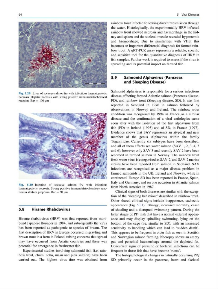
Fig. 5.29 Liver of sockeye salmon fry with infectious haematopoietic
necrosis. Hepatic necrosis with strong positive immunohistochemical
reaction. Bar ¼ 100 μm
Fig. 5.30 Intestine of sockeye salmon fry with infectious
haematopoietic necrosis. Strong positive immunohistochemistry reaction in stratum proprium. Bar ¼ 50 μm
64
5 Viral Diseases
Hirame Rhabdovirus
Hirame rhabdovirus (HRV) was first reported from moribund Japanese flounder in 1984, and subsequently the virus
has been reported as pathogenic to species of bream. The
first description of HRV in Europe occurred in grayling and
brown trout in a farm in Poland, raising concerns that spread
may have occurred from Asiatic countries and there was
potential for emergence in freshwater fish.
Experimental studies involving salmonid fish (i.e. rainbow trout, chum, coho, masu and pink salmon) have been
carried out. The highest virus titre was obtained from
rainbow trout infected following direct transmission through
the water. Histologically, the experimentally HRV infected
rainbow trout showed necrosis and haemorrhage in the kidney and spleen and the skeletal muscle revealed hyperaemia
and haemorrhage. Due to similarities with VHS, this
becomes an important differential diagnosis for farmed rainbow trout. A qRT-PCR assay represents a reliable, specific
and sensitive tool for the quantitative diagnosis of HRV in
fish samples. Further work is required to assess if the virus is
spreading and its potential impact on farmed fish.
5.9
Salmonid Alphavirus (Pancreas
and Sleeping Disease)
Salmonid alphavirus is responsible for a serious infectious
disease affecting farmed Atlantic salmon (Pancreas disease,
PD), and rainbow trout (Sleeping disease, SD). It was first
reported in Scotland in 1976 in salmon followed by
observations in Norway and Ireland. The rainbow trout
condition was recognised by 1994 in France as a similar
disease and the confirmation of a viral aetiologies came
soon after with the isolation of the first alphavirus from
fish (PD) in Ireland (1995) and of SD, in France (1997).
Evidence shows that SAV represents an atypical and new
member of the genus Alphavirus within the family
Togaviridae. Currently six subtypes have been described,
and all of them affects sea water salmon (SAV 1, 2, 3, 4, 5
and 6), however only SAV 3 and recently SAV 2 have been
recorded in farmed salmon in Norway. The rainbow trout
fresh water virus is categorised as SAV-2, and SAV-2 marine
strains have been reported from salmon in Scotland. SAV
infections are recognised as a major disease problem in
farmed salmonids in the UK, Ireland and Norway, while in
continental Europe SD has been reported in France, Spain,
Italy and Germany, and on one occasion in Atlantic salmon
from North America in 1987.
Clinical signs of both diseases are similar with the exception of the ‘sleeping behaviour’ described in rainbow trout.
Other shared clinical signs include inappetence, cachectic
appearance (Fig. 5.31), lethargy, increased mortality, cease
of shoaling and a disrupted swimming pattern. During the
latter stages of PD, fish that have a normal external appearance and may display spiralling swimming, lying on the
bottom of the cage (i.e. similar to SD), with an increased
sensitivity to handling which can lead to ‘sudden death’.
This appears to be frequent in older fish as seen in Scottish
and Norwegian salmon farming. Necropsy shows an empty
gut and petechial haemorrhage around the depleted fat.
Concurrent signs of parasitic or bacterial infections can be
frequent in those fish that have become ‘runts’.
The histopathological changes in naturally occurring PD/
SD primarily occur in the pancreas, heart and skeletal
Fig. 5.29 Liver of sockeye salmon fry with infectious haematopoietic
necrosis. Hepatic necrosis with strong positive immunohistochemical
reaction. Bar ¼ 100 μm
Fig. 5.30 Intestine of sockeye salmon fry with infectious
haematopoietic necrosis. Strong positive immunohistochemistry reaction in stratum proprium. Bar ¼ 50 μm
64
5 Viral Diseases