

Natural outbreaks of IHN are rare above 15
C and temperature is a factor which strongly influences disease progression. In acute infection mortality increases rapidly and fry
show lethargy and move to the edges of tanks. Abnormal
swimming patterns such as whirling and flashing may be
observed. Older fish rarely show behavioural changes. Pale
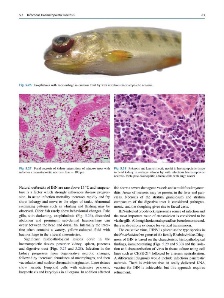
gills, skin darkening, exophthalmia (Fig. 5.26), distended
abdomen and prominent sub-dermal haemorrhage can
occur between the head and dorsal fin. Internally the intestine often contains a watery, yellow-coloured fluid with
haemorrhage in the visceral mesenteries.
Significant histopathological lesions occur in the
haematopoietic tissues, posterior kidney, spleen, pancreas
and digestive tract (Figs. 5.27 and 5.28). Infection in the
kidney progresses from degenerative necrotic changes,
followed by increased abundance of macrophages, and then
vacuolation and nuclear chromatin margination. Later tissues
show necrotic lymphoid cells with extensive pyknosis,
karyorrhexis and karyolysis in all organs. In addition affected
fish show a severe damage to vessels and a multifocal myocarditis. Areas of necrosis may be present in the liver and pancreas. Necrosis of the stratum granulosum and stratum
compactum of the digestive tract is considered pathognomonic, and the sloughing gives rise to faecal casts.
IHN-infected broodstock represent a source of infection and
the most important route of transmission is considered to be
via the gills. Although horizontal spread has been demonstrated,
there is also strong evidence for vertical transmission.
The causative virus, IHNV is placed as the type species in
the Novirhabdovirus genus of the family Rhabdoviridae. Diagnosis of IHN is based on the characteristic histopathological
findings, immunostaining (Figs. 5.29 and 5.30) and the isolation and characterisation of virus in tissue culture using cell
lines such as CHSE-214 followed by a serum neutralisation.
A differential diagnosis would include infectious pancreatic
necrosis. There is evidence that an orally delivered DNA
vaccine for IHN is achievable, but this approach requires
refinement.
Fig. 5.26 Exopthalmia with haemorrhage in rainbow trout fry with infectious haematopoietic necrosis
Fig. 5.27 Focal necrosis of kidney interstitium of rainbow trout with
infectious haematopoietic necrosis. Bar ¼ 100 μm
Fig. 5.28 Pyknotic and karryorrhectic nuclei in haematopoietic tissue
in head kidney in sockeye salmon fry with infectious haematopoietic
necrosis. Note pale eosinophilic adrenal cells with large nuclei
5.7 Infectious Haematopoietic Necrosis
63
C and temperature is a factor which strongly influences disease progression. In acute infection mortality increases rapidly and fry
show lethargy and move to the edges of tanks. Abnormal
swimming patterns such as whirling and flashing may be
observed. Older fish rarely show behavioural changes. Pale
gills, skin darkening, exophthalmia (Fig. 5.26), distended
abdomen and prominent sub-dermal haemorrhage can
occur between the head and dorsal fin. Internally the intestine often contains a watery, yellow-coloured fluid with
haemorrhage in the visceral mesenteries.
Significant histopathological lesions occur in the
haematopoietic tissues, posterior kidney, spleen, pancreas
and digestive tract (Figs. 5.27 and 5.28). Infection in the
kidney progresses from degenerative necrotic changes,
followed by increased abundance of macrophages, and then
vacuolation and nuclear chromatin margination. Later tissues
show necrotic lymphoid cells with extensive pyknosis,
karyorrhexis and karyolysis in all organs. In addition affected
fish show a severe damage to vessels and a multifocal myocarditis. Areas of necrosis may be present in the liver and pancreas. Necrosis of the stratum granulosum and stratum
compactum of the digestive tract is considered pathognomonic, and the sloughing gives rise to faecal casts.
IHN-infected broodstock represent a source of infection and
the most important route of transmission is considered to be
via the gills. Although horizontal spread has been demonstrated,
there is also strong evidence for vertical transmission.
The causative virus, IHNV is placed as the type species in
the Novirhabdovirus genus of the family Rhabdoviridae. Diagnosis of IHN is based on the characteristic histopathological
findings, immunostaining (Figs. 5.29 and 5.30) and the isolation and characterisation of virus in tissue culture using cell
lines such as CHSE-214 followed by a serum neutralisation.
A differential diagnosis would include infectious pancreatic
necrosis. There is evidence that an orally delivered DNA
vaccine for IHN is achievable, but this approach requires
refinement.
Fig. 5.26 Exopthalmia with haemorrhage in rainbow trout fry with infectious haematopoietic necrosis
Fig. 5.27 Focal necrosis of kidney interstitium of rainbow trout with
infectious haematopoietic necrosis. Bar ¼ 100 μm
Fig. 5.28 Pyknotic and karryorrhectic nuclei in haematopoietic tissue
in head kidney in sockeye salmon fry with infectious haematopoietic
necrosis. Note pale eosinophilic adrenal cells with large nuclei
5.7 Infectious Haematopoietic Necrosis
63