

muscle, although the severity and the distribution of the
lesions depend on the time after initial infection. As infection may occur asynchronously within the same population
in a cage, it is common to find fish from the same unit
displaying different levels and severity of pathological
changes. The first lesions are an acute phase of pancreatic
acinar cell necrosis, with variable inflammatory response
spanning from virtually none to a moderate mononuclear
cell infiltration and/or fibrosis of the periacinar tissue, with
loss of exocrine tissue (Fig. 5.32). The endocrine pancreas is
not a target tissue. Almost simultaneously or slightly
delayed, heart lesions can be observed. A severe degeneration with multifocal cardiomyocytic necrosis affecting the
spongy and compact myocardium is described. Lesions are
characterised by individual or clusters of shrunken cells
becoming strongly eosinophilic with pyknotic nuclei.
Increased cellularity at the junction of the ventricle compact
–spongy layer can be observed and hypertrophy of
cardiomyocytic nuclei also described, the latter being evident in the recovery phase (Figs. 5.33 and 5.34). A distinct
difference in the response of affected fish in relation to their
life stage (smolt versus growers) has been observed, with
mitotic figures being reported as a consistent feature in the
heart of affected smolts, but less frequently or absent in older
fish. Finally, the skeletal muscle becomes involved at least
3–4 weeks after the lesions in the pancreas and heart are
observed. Both red and white muscle are affected and
characterised by myofibre hyaline degeneration with
swollen and fragmented sarcoplasm (Fig. 5.35). In the
white muscle individual fibres are affected and can be seen
as highly eosinophilic with central migration of nuclei. Sarcoplasm infiltration by phagocytic macrophages and at later
stage, a variable inflammation and fibrosis can be noted. The
red muscle layer shows similar sarcoplasmic pathological
changes, although frequently the proportion of damaged
tissue is greater than in the white muscle. A variable degree
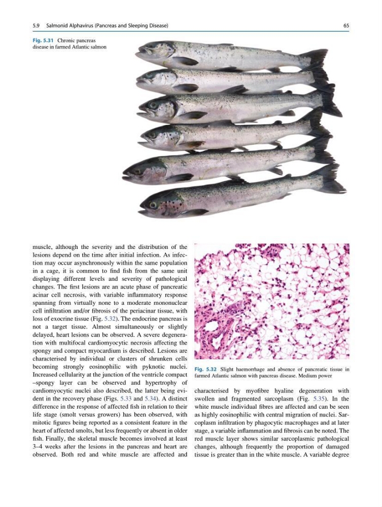
Fig. 5.31 Chronic pancreas
disease in farmed Atlantic salmon
Fig. 5.32 Slight haemorrhage and absence of pancreatic tissue in
farmed Atlantic salmon with pancreas disease. Medium power
5.9 Salmonid Alphavirus (Pancreas and Sleeping Disease)
65
lesions depend on the time after initial infection. As infection may occur asynchronously within the same population
in a cage, it is common to find fish from the same unit
displaying different levels and severity of pathological
changes. The first lesions are an acute phase of pancreatic
acinar cell necrosis, with variable inflammatory response
spanning from virtually none to a moderate mononuclear
cell infiltration and/or fibrosis of the periacinar tissue, with
loss of exocrine tissue (Fig. 5.32). The endocrine pancreas is
not a target tissue. Almost simultaneously or slightly
delayed, heart lesions can be observed. A severe degeneration with multifocal cardiomyocytic necrosis affecting the
spongy and compact myocardium is described. Lesions are
characterised by individual or clusters of shrunken cells
becoming strongly eosinophilic with pyknotic nuclei.
Increased cellularity at the junction of the ventricle compact
–spongy layer can be observed and hypertrophy of
cardiomyocytic nuclei also described, the latter being evident in the recovery phase (Figs. 5.33 and 5.34). A distinct
difference in the response of affected fish in relation to their
life stage (smolt versus growers) has been observed, with
mitotic figures being reported as a consistent feature in the
heart of affected smolts, but less frequently or absent in older
fish. Finally, the skeletal muscle becomes involved at least
3–4 weeks after the lesions in the pancreas and heart are
observed. Both red and white muscle are affected and
characterised by myofibre hyaline degeneration with
swollen and fragmented sarcoplasm (Fig. 5.35). In the
white muscle individual fibres are affected and can be seen
as highly eosinophilic with central migration of nuclei. Sarcoplasm infiltration by phagocytic macrophages and at later
stage, a variable inflammation and fibrosis can be noted. The
red muscle layer shows similar sarcoplasmic pathological
changes, although frequently the proportion of damaged
tissue is greater than in the white muscle. A variable degree
Fig. 5.31 Chronic pancreas
disease in farmed Atlantic salmon
Fig. 5.32 Slight haemorrhage and absence of pancreatic tissue in
farmed Atlantic salmon with pancreas disease. Medium power
5.9 Salmonid Alphavirus (Pancreas and Sleeping Disease)
65