

may become infected in fresh and marine environments. The
emergence of VHSV in the Great Lakes Basin of North
America during 2003 highlighted this virus as a cause or
mortality in a range of freshwater fish species. Outbreaks
typically result in an acute to chronic disease when
temperatures are fluctuating and generally below 14
C.
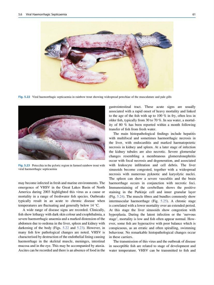
A wide range of disease signs are recorded. Clinically,
fish show lethargy with dark skin colour and exophthalmia, a
severe haemorrhagic anaemia and a marked distension of the
abdomen due to oedema in the liver, spleen and kidney with
darkening of the body (Figs. 5.22 and 5.23). However, in
many fish few pathological changes are noted. VHSV is
characterised by destruction of the endothelial lining causing
haemorrhage in the skeletal muscle, meninges, intestinal
mucosa and in the eye. This may be accompanied by ataxia.
Ascites can be recorded and there is an absence of food in the
gastrointestinal tract. These acute signs are usually
associated with a rapid onset of heavy mortality and linked
to the age of the fish with up to 100 % in fry, often less in
older fish, typically from 30 to 70 %. In sea water, a mortality of 80 % has been reported within a month following
transfer of fish from fresh water.
The main histopathological findings include hepatitis
with multifocal and sometimes haemorrhagic necrosis in
the liver, with endocarditis and marked haematopoietic
necrosis in kidney and spleen. At a later stage of infection
the kidney tubules are also necrotic. Severe glomerular
changes resembling a membranous glomerulonephritis
occur with focal necrosis and degeneration, and associated
with leukocyte infiltration and cell debris. The liver
sinusoids become congested, together with a widespread
necrosis with numerous pyknotic and karyolytic nuclei.
The spleen can show a severe vasculitis and the brain
haemorrhage occurs in conjunction with necrotic foci.
Immunostaining of the cerebellum shows the positive
staining in the Purkinje cell and inner granular layer
(Fig. 5.24). The muscle fibres and bundles commonly show
intermuscular haemorrhage (Fig. 5.25). A chronic stage
is correlated with a lower mortality over an extended period.
At this stage the liver sinusoids show congestion with
hyperplasia. During the latent infection or the ‘nervous
stage’, mortality is low and fish often appear normal. However, some fish are hyperactive with poor balance which is
conspicuous, as an erratic and often spiralling, swimming
behaviour. No remarkable histopathological changes occur
in these carriers.
The transmission of this virus and the outbreak of disease
in susceptible fish are related to stage of development and
water temperature. VHSV can be transmitted to fish and
Fig. 5.22 Viral haemorrhagic septicaemia in rainbow trout showing widespread petechiae of the musculature and pale gills
Fig. 5.23 Petecchia in the pyloric region in farmed rainbow trout with
viral haemorrhagic septicaemia
5.6 Viral Haemorrhagic Septicaemia
61
emergence of VHSV in the Great Lakes Basin of North
America during 2003 highlighted this virus as a cause or
mortality in a range of freshwater fish species. Outbreaks
typically result in an acute to chronic disease when
temperatures are fluctuating and generally below 14
C.
A wide range of disease signs are recorded. Clinically,
fish show lethargy with dark skin colour and exophthalmia, a
severe haemorrhagic anaemia and a marked distension of the
abdomen due to oedema in the liver, spleen and kidney with
darkening of the body (Figs. 5.22 and 5.23). However, in
many fish few pathological changes are noted. VHSV is
characterised by destruction of the endothelial lining causing
haemorrhage in the skeletal muscle, meninges, intestinal
mucosa and in the eye. This may be accompanied by ataxia.
Ascites can be recorded and there is an absence of food in the
gastrointestinal tract. These acute signs are usually
associated with a rapid onset of heavy mortality and linked
to the age of the fish with up to 100 % in fry, often less in
older fish, typically from 30 to 70 %. In sea water, a mortality of 80 % has been reported within a month following
transfer of fish from fresh water.
The main histopathological findings include hepatitis
with multifocal and sometimes haemorrhagic necrosis in
the liver, with endocarditis and marked haematopoietic
necrosis in kidney and spleen. At a later stage of infection
the kidney tubules are also necrotic. Severe glomerular
changes resembling a membranous glomerulonephritis
occur with focal necrosis and degeneration, and associated
with leukocyte infiltration and cell debris. The liver
sinusoids become congested, together with a widespread
necrosis with numerous pyknotic and karyolytic nuclei.
The spleen can show a severe vasculitis and the brain
haemorrhage occurs in conjunction with necrotic foci.
Immunostaining of the cerebellum shows the positive
staining in the Purkinje cell and inner granular layer
(Fig. 5.24). The muscle fibres and bundles commonly show
intermuscular haemorrhage (Fig. 5.25). A chronic stage
is correlated with a lower mortality over an extended period.
At this stage the liver sinusoids show congestion with
hyperplasia. During the latent infection or the ‘nervous
stage’, mortality is low and fish often appear normal. However, some fish are hyperactive with poor balance which is
conspicuous, as an erratic and often spiralling, swimming
behaviour. No remarkable histopathological changes occur
in these carriers.
The transmission of this virus and the outbreak of disease
in susceptible fish are related to stage of development and
water temperature. VHSV can be transmitted to fish and
Fig. 5.22 Viral haemorrhagic septicaemia in rainbow trout showing widespread petechiae of the musculature and pale gills
Fig. 5.23 Petecchia in the pyloric region in farmed rainbow trout with
viral haemorrhagic septicaemia
5.6 Viral Haemorrhagic Septicaemia
61