

5.4
Piscine Reovirus (Heart and Skeletal
Muscle Inflammation, HSMI)
Piscine reovirus (PRV) has been recently reported as the
aetiological agent of HSMI, a systemic viral disease of seawater farmed Atlantic salmon. The first cases were identified
in Norway in 1999 and the disease is currently widespread in
Norwegian aquaculture where it causes substantial losses.
This condition has also been described from farmed salmon
in Scotland. The PCR-screening of marine fish caught along
the Norwegian coast has revealed PRV in great silver smelt,
capelin, Atlantic herring and horse mackerel.
Clinical outbreaks typically occur 5–9 months after transfer to sea water. Morbidity may be very high in affected
cages, while mortality may reach 20 %. Clinical signs
include anorexia and abnormal swimming behaviour and
internally, pale heart, yellow-orange liver, ascites, splenomegaly and visceral petechiae.
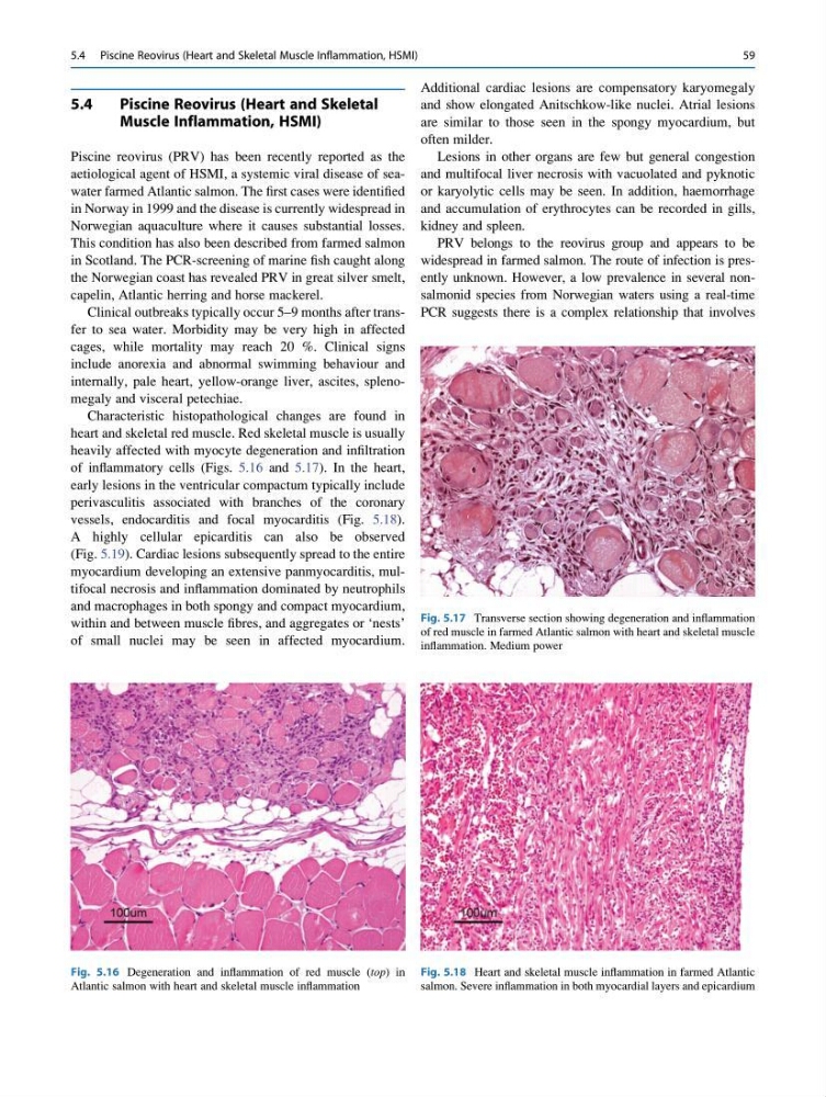
Characteristic histopathological changes are found in
heart and skeletal red muscle. Red skeletal muscle is usually
heavily affected with myocyte degeneration and infiltration
of inflammatory cells (Figs. 5.16 and 5.17). In the heart,
early lesions in the ventricular compactum typically include
perivasculitis associated with branches of the coronary
vessels, endocarditis and focal myocarditis (Fig. 5.18).
A highly cellular epicarditis can also be observed
(Fig. 5.19). Cardiac lesions subsequently spread to the entire
myocardium developing an extensive panmyocarditis, multifocal necrosis and inflammation dominated by neutrophils
and macrophages in both spongy and compact myocardium,
within and between muscle fibres, and aggregates or ‘nests’
of small nuclei may be seen in affected myocardium.
Additional cardiac lesions are compensatory karyomegaly
and show elongated Anitschkow-like nuclei. Atrial lesions
are similar to those seen in the spongy myocardium, but
often milder.
Lesions in other organs are few but general congestion
and multifocal liver necrosis with vacuolated and pyknotic
or karyolytic cells may be seen. In addition, haemorrhage
and accumulation of erythrocytes can be recorded in gills,
kidney and spleen.
PRV belongs to the reovirus group and appears to be
widespread in farmed salmon. The route of infection is presently unknown. However, a low prevalence in several nonsalmonid species from Norwegian waters using a real-time
PCR suggests there is a complex relationship that involves
Fig. 5.16 Degeneration and inflammation of red muscle (top) in
Atlantic salmon with heart and skeletal muscle inflammation
Fig. 5.17 Transverse section showing degeneration and inflammation
of red muscle in farmed Atlantic salmon with heart and skeletal muscle
inflammation. Medium power
Fig. 5.18 Heart and skeletal muscle inflammation in farmed Atlantic
salmon. Severe inflammation in both myocardial layers and epicardium
5.4 Piscine Reovirus (Heart and Skeletal Muscle Inflammation, HSMI)
59
Piscine Reovirus (Heart and Skeletal
Muscle Inflammation, HSMI)
Piscine reovirus (PRV) has been recently reported as the
aetiological agent of HSMI, a systemic viral disease of seawater farmed Atlantic salmon. The first cases were identified
in Norway in 1999 and the disease is currently widespread in
Norwegian aquaculture where it causes substantial losses.
This condition has also been described from farmed salmon
in Scotland. The PCR-screening of marine fish caught along
the Norwegian coast has revealed PRV in great silver smelt,
capelin, Atlantic herring and horse mackerel.
Clinical outbreaks typically occur 5–9 months after transfer to sea water. Morbidity may be very high in affected
cages, while mortality may reach 20 %. Clinical signs
include anorexia and abnormal swimming behaviour and
internally, pale heart, yellow-orange liver, ascites, splenomegaly and visceral petechiae.
Characteristic histopathological changes are found in
heart and skeletal red muscle. Red skeletal muscle is usually
heavily affected with myocyte degeneration and infiltration
of inflammatory cells (Figs. 5.16 and 5.17). In the heart,
early lesions in the ventricular compactum typically include
perivasculitis associated with branches of the coronary
vessels, endocarditis and focal myocarditis (Fig. 5.18).
A highly cellular epicarditis can also be observed
(Fig. 5.19). Cardiac lesions subsequently spread to the entire
myocardium developing an extensive panmyocarditis, multifocal necrosis and inflammation dominated by neutrophils
and macrophages in both spongy and compact myocardium,
within and between muscle fibres, and aggregates or ‘nests’
of small nuclei may be seen in affected myocardium.
Additional cardiac lesions are compensatory karyomegaly
and show elongated Anitschkow-like nuclei. Atrial lesions
are similar to those seen in the spongy myocardium, but
often milder.
Lesions in other organs are few but general congestion
and multifocal liver necrosis with vacuolated and pyknotic
or karyolytic cells may be seen. In addition, haemorrhage
and accumulation of erythrocytes can be recorded in gills,
kidney and spleen.
PRV belongs to the reovirus group and appears to be
widespread in farmed salmon. The route of infection is presently unknown. However, a low prevalence in several nonsalmonid species from Norwegian waters using a real-time
PCR suggests there is a complex relationship that involves
Fig. 5.16 Degeneration and inflammation of red muscle (top) in
Atlantic salmon with heart and skeletal muscle inflammation
Fig. 5.17 Transverse section showing degeneration and inflammation
of red muscle in farmed Atlantic salmon with heart and skeletal muscle
inflammation. Medium power
Fig. 5.18 Heart and skeletal muscle inflammation in farmed Atlantic
salmon. Severe inflammation in both myocardial layers and epicardium
5.4 Piscine Reovirus (Heart and Skeletal Muscle Inflammation, HSMI)
59