

genetically differentiated on the basis of the sequence of a
highly polymorphic region (HPR of genomic segment 6
which encodes the Haemagglutinin-Esterase (HE) protein).
A deletion within the HPR region (named HPRΔ ISAV)
in certain ISAV variants appears to be a dependable indicator of pathogenicity. ISAV without any deletions in the
HPR region (HPR0 ISAV) has been reported only in apparently healthy fish and to date have not been associated
with ISA disease. A reverse transcriptase-polymerase
chain reaction has been developed as a sensitive method
to detect carrier fish. A commercial vaccine is also
available.
5.3
Oncorhynchus masou Virus
Oncorhynchus masou virus (OMV) is a virulent and economical significant disease that was originally isolated from
ovarian fluids of a landlocked population of adult masou
salmon in Hokkaido, Japan, but now also occurring in wild
stocks. Other salmonid species are susceptible to OMV
including coho, chum, kokanee and rainbow trout with
high mortality in young fish.
Affected fish are dark, frequently showing severe
exophthalmia and petechial haemorrhage under the lower
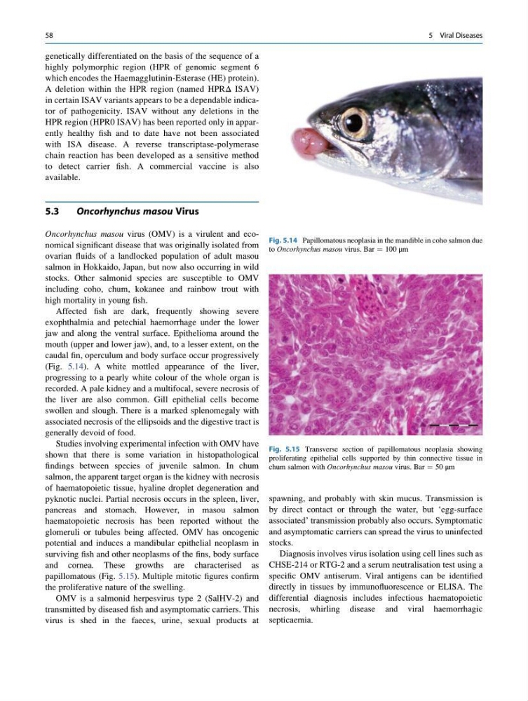
jaw and along the ventral surface. Epithelioma around the
mouth (upper and lower jaw), and, to a lesser extent, on the
caudal fin, operculum and body surface occur progressively
(Fig. 5.14). A white mottled appearance of the liver,
progressing to a pearly white colour of the whole organ is
recorded. A pale kidney and a multifocal, severe necrosis of
the liver are also common. Gill epithelial cells become
swollen and slough. There is a marked splenomegaly with
associated necrosis of the ellipsoids and the digestive tract is
generally devoid of food.
Studies involving experimental infection with OMV have
shown that there is some variation in histopathological
findings between species of juvenile salmon. In chum
salmon, the apparent target organ is the kidney with necrosis
of haematopoietic tissue, hyaline droplet degeneration and
pyknotic nuclei. Partial necrosis occurs in the spleen, liver,
pancreas and stomach. However, in masou salmon
haematopoietic necrosis has been reported without the
glomeruli or tubules being affected. OMV has oncogenic
potential and induces a mandibular epithelial neoplasm in
surviving fish and other neoplasms of the fins, body surface
and cornea. These growths are characterised as
papillomatous (Fig. 5.15). Multiple mitotic figures confirm
the proliferative nature of the swelling.
OMV is a salmonid herpesvirus type 2 (SalHV-2) and
transmitted by diseased fish and asymptomatic carriers. This
virus is shed in the faeces, urine, sexual products at
spawning, and probably with skin mucus. Transmission is
by direct contact or through the water, but ‘egg-surface
associated’ transmission probably also occurs. Symptomatic
and asymptomatic carriers can spread the virus to uninfected
stocks.
Diagnosis involves virus isolation using cell lines such as
CHSE-214 or RTG-2 and a serum neutralisation test using a
specific OMV antiserum. Viral antigens can be identified
directly in tissues by immunofluorescence or ELISA. The
differential diagnosis includes infectious haematopoietic
necrosis, whirling disease and viral haemorrhagic
septicaemia.
Fig. 5.14 Papillomatous neoplasia in the mandible in coho salmon due
to Oncorhynchus masou virus. Bar ¼ 100 μm
Fig. 5.15 Transverse section of papillomatous neoplasia showing
proliferating epithelial cells supported by thin connective tissue in
chum salmon with Oncorhynchus masou virus. Bar ¼ 50 μm
58
5 Viral Diseases
highly polymorphic region (HPR of genomic segment 6
which encodes the Haemagglutinin-Esterase (HE) protein).
A deletion within the HPR region (named HPRΔ ISAV)
in certain ISAV variants appears to be a dependable indicator of pathogenicity. ISAV without any deletions in the
HPR region (HPR0 ISAV) has been reported only in apparently healthy fish and to date have not been associated
with ISA disease. A reverse transcriptase-polymerase
chain reaction has been developed as a sensitive method
to detect carrier fish. A commercial vaccine is also
available.
5.3
Oncorhynchus masou Virus
Oncorhynchus masou virus (OMV) is a virulent and economical significant disease that was originally isolated from
ovarian fluids of a landlocked population of adult masou
salmon in Hokkaido, Japan, but now also occurring in wild
stocks. Other salmonid species are susceptible to OMV
including coho, chum, kokanee and rainbow trout with
high mortality in young fish.
Affected fish are dark, frequently showing severe
exophthalmia and petechial haemorrhage under the lower
jaw and along the ventral surface. Epithelioma around the
mouth (upper and lower jaw), and, to a lesser extent, on the
caudal fin, operculum and body surface occur progressively
(Fig. 5.14). A white mottled appearance of the liver,
progressing to a pearly white colour of the whole organ is
recorded. A pale kidney and a multifocal, severe necrosis of
the liver are also common. Gill epithelial cells become
swollen and slough. There is a marked splenomegaly with
associated necrosis of the ellipsoids and the digestive tract is
generally devoid of food.
Studies involving experimental infection with OMV have
shown that there is some variation in histopathological
findings between species of juvenile salmon. In chum
salmon, the apparent target organ is the kidney with necrosis
of haematopoietic tissue, hyaline droplet degeneration and
pyknotic nuclei. Partial necrosis occurs in the spleen, liver,
pancreas and stomach. However, in masou salmon
haematopoietic necrosis has been reported without the
glomeruli or tubules being affected. OMV has oncogenic
potential and induces a mandibular epithelial neoplasm in
surviving fish and other neoplasms of the fins, body surface
and cornea. These growths are characterised as
papillomatous (Fig. 5.15). Multiple mitotic figures confirm
the proliferative nature of the swelling.
OMV is a salmonid herpesvirus type 2 (SalHV-2) and
transmitted by diseased fish and asymptomatic carriers. This
virus is shed in the faeces, urine, sexual products at
spawning, and probably with skin mucus. Transmission is
by direct contact or through the water, but ‘egg-surface
associated’ transmission probably also occurs. Symptomatic
and asymptomatic carriers can spread the virus to uninfected
stocks.
Diagnosis involves virus isolation using cell lines such as
CHSE-214 or RTG-2 and a serum neutralisation test using a
specific OMV antiserum. Viral antigens can be identified
directly in tissues by immunofluorescence or ELISA. The
differential diagnosis includes infectious haematopoietic
necrosis, whirling disease and viral haemorrhagic
septicaemia.
Fig. 5.14 Papillomatous neoplasia in the mandible in coho salmon due
to Oncorhynchus masou virus. Bar ¼ 100 μm
Fig. 5.15 Transverse section of papillomatous neoplasia showing
proliferating epithelial cells supported by thin connective tissue in
chum salmon with Oncorhynchus masou virus. Bar ¼ 50 μm
58
5 Viral Diseases