

complete loss of all histological features. Overall, necrotic
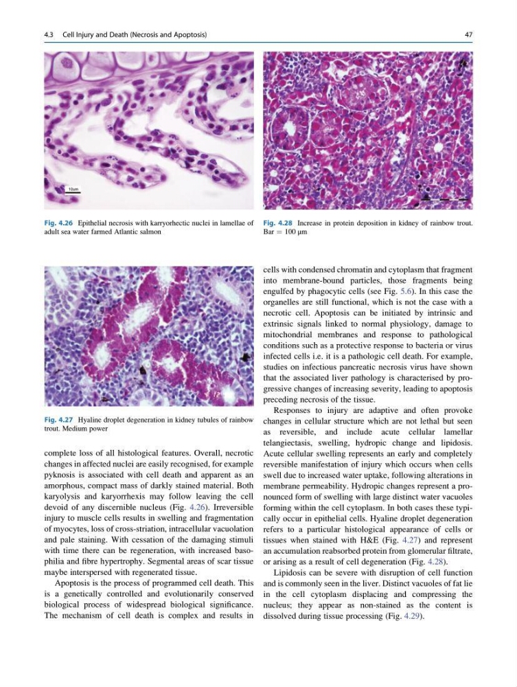
changes in affected nuclei are easily recognised, for example
pyknosis is associated with cell death and apparent as an
amorphous, compact mass of darkly stained material. Both
karyolysis and karyorrhexis may follow leaving the cell
devoid of any discernible nucleus (Fig. 4.26). Irreversible
injury to muscle cells results in swelling and fragmentation
of myocytes, loss of cross-striation, intracellular vacuolation
and pale staining. With cessation of the damaging stimuli
with time there can be regeneration, with increased basophilia and fibre hypertrophy. Segmental areas of scar tissue
maybe interspersed with regenerated tissue.
Apoptosis is the process of programmed cell death. This
is a genetically controlled and evolutionarily conserved
biological process of widespread biological significance.
The mechanism of cell death is complex and results in
cells with condensed chromatin and cytoplasm that fragment
into membrane-bound particles, those fragments being
engulfed by phagocytic cells (see Fig. 5.6). In this case the
organelles are still functional, which is not the case with a
necrotic cell. Apoptosis can be initiated by intrinsic and
extrinsic signals linked to normal physiology, damage to
mitochondrial membranes and response to pathological
conditions such as a protective response to bacteria or virus
infected cells i.e. it is a pathologic cell death. For example,
studies on infectious pancreatic necrosis virus have shown
that the associated liver pathology is characterised by progressive changes of increasing severity, leading to apoptosis
preceding necrosis of the tissue.
Responses to injury are adaptive and often provoke
changes in cellular structure which are not lethal but seen
as reversible, and include acute cellular lamellar
telangiectasis, swelling, hydropic change and lipidosis.
Acute cellular swelling represents an early and completely
reversible manifestation of injury which occurs when cells
swell due to increased water uptake, following alterations in
membrane permeability. Hydropic changes represent a pronounced form of swelling with large distinct water vacuoles
forming within the cell cytoplasm. In both cases these typically occur in epithelial cells. Hyaline droplet degeneration
refers to a particular histological appearance of cells or
tissues when stained with H&E (Fig. 4.27) and represent
an accumulation reabsorbed protein from glomerular filtrate,
or arising as a result of cell degeneration (Fig. 4.28).
Lipidosis can be severe with disruption of cell function
and is commonly seen in the liver. Distinct vacuoles of fat lie
in the cell cytoplasm displacing and compressing the
nucleus; they appear as non-stained as the content is
dissolved during tissue processing (Fig. 4.29).
Fig. 4.26 Epithelial necrosis with karryorhectic nuclei in lamellae of
adult sea water farmed Atlantic salmon
Fig. 4.27 Hyaline droplet degeneration in kidney tubules of rainbow
trout. Medium power
Fig. 4.28 Increase in protein deposition in kidney of rainbow trout.
Bar ¼ 100 μm
4.3 Cell Injury and Death (Necrosis and Apoptosis)
47
changes in affected nuclei are easily recognised, for example
pyknosis is associated with cell death and apparent as an
amorphous, compact mass of darkly stained material. Both
karyolysis and karyorrhexis may follow leaving the cell
devoid of any discernible nucleus (Fig. 4.26). Irreversible
injury to muscle cells results in swelling and fragmentation
of myocytes, loss of cross-striation, intracellular vacuolation
and pale staining. With cessation of the damaging stimuli
with time there can be regeneration, with increased basophilia and fibre hypertrophy. Segmental areas of scar tissue
maybe interspersed with regenerated tissue.
Apoptosis is the process of programmed cell death. This
is a genetically controlled and evolutionarily conserved
biological process of widespread biological significance.
The mechanism of cell death is complex and results in
cells with condensed chromatin and cytoplasm that fragment
into membrane-bound particles, those fragments being
engulfed by phagocytic cells (see Fig. 5.6). In this case the
organelles are still functional, which is not the case with a
necrotic cell. Apoptosis can be initiated by intrinsic and
extrinsic signals linked to normal physiology, damage to
mitochondrial membranes and response to pathological
conditions such as a protective response to bacteria or virus
infected cells i.e. it is a pathologic cell death. For example,
studies on infectious pancreatic necrosis virus have shown
that the associated liver pathology is characterised by progressive changes of increasing severity, leading to apoptosis
preceding necrosis of the tissue.
Responses to injury are adaptive and often provoke
changes in cellular structure which are not lethal but seen
as reversible, and include acute cellular lamellar
telangiectasis, swelling, hydropic change and lipidosis.
Acute cellular swelling represents an early and completely
reversible manifestation of injury which occurs when cells
swell due to increased water uptake, following alterations in
membrane permeability. Hydropic changes represent a pronounced form of swelling with large distinct water vacuoles
forming within the cell cytoplasm. In both cases these typically occur in epithelial cells. Hyaline droplet degeneration
refers to a particular histological appearance of cells or
tissues when stained with H&E (Fig. 4.27) and represent
an accumulation reabsorbed protein from glomerular filtrate,
or arising as a result of cell degeneration (Fig. 4.28).
Lipidosis can be severe with disruption of cell function
and is commonly seen in the liver. Distinct vacuoles of fat lie
in the cell cytoplasm displacing and compressing the
nucleus; they appear as non-stained as the content is
dissolved during tissue processing (Fig. 4.29).
Fig. 4.26 Epithelial necrosis with karryorhectic nuclei in lamellae of
adult sea water farmed Atlantic salmon
Fig. 4.27 Hyaline droplet degeneration in kidney tubules of rainbow
trout. Medium power
Fig. 4.28 Increase in protein deposition in kidney of rainbow trout.
Bar ¼ 100 μm
4.3 Cell Injury and Death (Necrosis and Apoptosis)
47