

An excessive amount of fluid in or around cells, tissues or
serous cavities is termed oedema and may follow changes in
hydrostatic or osmotic pressure and increases in vascular permeability that accompany inflammation, as seen in fish infected
with Aliivibrio salmonicida. For example, eye oedema can
occur in the cornea, retina and choroid rete mirabile. Corneal
oedema typically accompanies eye disease or wounding and
follows separation of the collagen fibres and Malpighian cells of
the stroma resulting in a hazy or cloudy appearance. Extensive
fibrinous exudates, are often infiltrated with neutrophils, however these are common to a number of pathological conditions.
Hepatocellular hydropic degeneration can be associated with
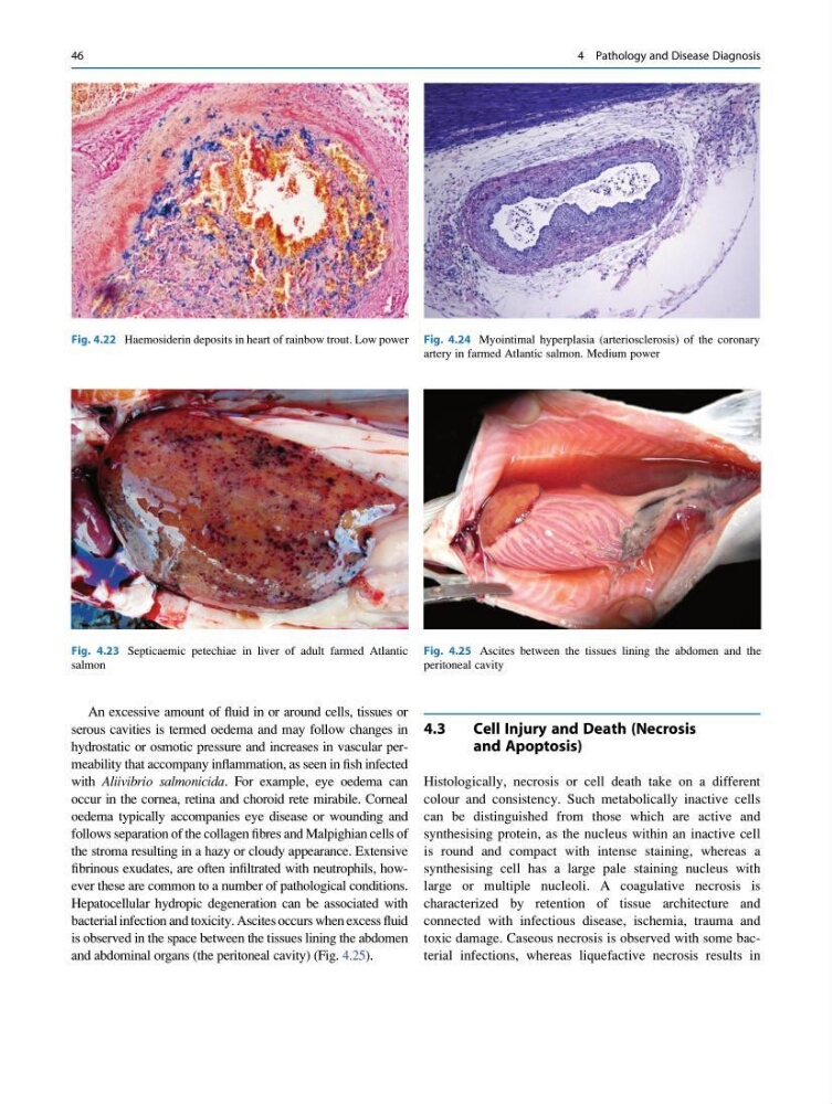
bacterial infection and toxicity. Ascites occurs when excess fluid
is observed in the space between the tissues lining the abdomen
and abdominal organs (the peritoneal cavity) (Fig. 4.25).
4.3
Cell Injury and Death (Necrosis
and Apoptosis)
Histologically, necrosis or cell death take on a different
colour and consistency. Such metabolically inactive cells
can be distinguished from those which are active and
synthesising protein, as the nucleus within an inactive cell
is round and compact with intense staining, whereas a
synthesising cell has a large pale staining nucleus with
large or multiple nucleoli. A coagulative necrosis is
characterized by retention of tissue architecture and
connected with infectious disease, ischemia, trauma and
toxic damage. Caseous necrosis is observed with some bacterial infections, whereas liquefactive necrosis results in
Fig. 4.23 Septicaemic petechiae in liver of adult farmed Atlantic
salmon
Fig. 4.24 Myointimal hyperplasia (arteriosclerosis) of the coronary
artery in farmed Atlantic salmon. Medium power
Fig. 4.25 Ascites between the tissues lining the abdomen and the
peritoneal cavity
Fig. 4.22 Haemosiderin deposits in heart of rainbow trout. Low power
46
4 Pathology and Disease Diagnosis
serous cavities is termed oedema and may follow changes in
hydrostatic or osmotic pressure and increases in vascular permeability that accompany inflammation, as seen in fish infected
with Aliivibrio salmonicida. For example, eye oedema can
occur in the cornea, retina and choroid rete mirabile. Corneal
oedema typically accompanies eye disease or wounding and
follows separation of the collagen fibres and Malpighian cells of
the stroma resulting in a hazy or cloudy appearance. Extensive
fibrinous exudates, are often infiltrated with neutrophils, however these are common to a number of pathological conditions.
Hepatocellular hydropic degeneration can be associated with
bacterial infection and toxicity. Ascites occurs when excess fluid
is observed in the space between the tissues lining the abdomen
and abdominal organs (the peritoneal cavity) (Fig. 4.25).
4.3
Cell Injury and Death (Necrosis
and Apoptosis)
Histologically, necrosis or cell death take on a different
colour and consistency. Such metabolically inactive cells
can be distinguished from those which are active and
synthesising protein, as the nucleus within an inactive cell
is round and compact with intense staining, whereas a
synthesising cell has a large pale staining nucleus with
large or multiple nucleoli. A coagulative necrosis is
characterized by retention of tissue architecture and
connected with infectious disease, ischemia, trauma and
toxic damage. Caseous necrosis is observed with some bacterial infections, whereas liquefactive necrosis results in
Fig. 4.23 Septicaemic petechiae in liver of adult farmed Atlantic
salmon
Fig. 4.24 Myointimal hyperplasia (arteriosclerosis) of the coronary
artery in farmed Atlantic salmon. Medium power
Fig. 4.25 Ascites between the tissues lining the abdomen and the
peritoneal cavity
Fig. 4.22 Haemosiderin deposits in heart of rainbow trout. Low power
46
4 Pathology and Disease Diagnosis