

on the ventral parts of the fish (Fig. 4.23). A haemorrhagic
diathesis in the liver is reported for Atlantic salmon smolts
suffering from haemorrhagic smolt syndrome (see
Figs. 11.16 and 11.17).
Hyperaemia and congestion originate from locally
increased blood volumes. Hyperaemia is an active process
and implies arterial side engorgement of the vascular bed,
while congestion indicates a passive process resulting from
reduced outflow of blood from a tissue. Hyperaemia is
usually accompanied by evidence of inflammation, and is
associated with vascular dilation due to localized release of
inflammatory mediators. Passive congestion is linked with
reduction in venous outflow due to non-inflammatory
events such as cardiac failure, constriction or obstruction
of vascular outflow due to tissue torsion, neoplasia, or other
compressive events. It is often difficult to distinguish
hyperaemia from congestion. Fish can show a number of
cardiovascular changes when exposed to hypoxia as result
of depletion of oxygen in the water, and ultimately, this
leads to vascular congestion and necrotic or apoptotic
lesions.
Myointimal hyperplasia (arteriosclerosis) is usually restricted
to the main coronary artery and occurs progressively in the
majority of salmonids during sexual maturation and at spawning
(Fig. 4.24). The initiating mechanism for coronary lesion formation appears to be vascular injury to the coronary artery, as a result
of the bulbus arteriosus being markedly distended.
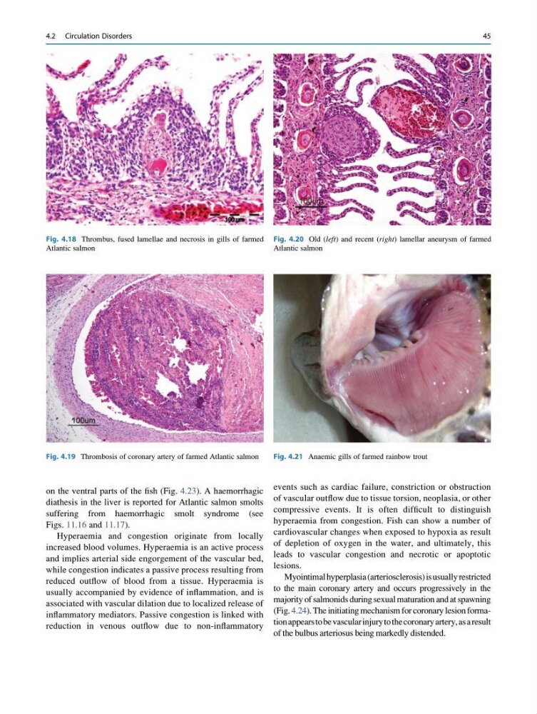
Fig. 4.18 Thrombus, fused lamellae and necrosis in gills of farmed
Atlantic salmon
Fig. 4.19 Thrombosis of coronary artery of farmed Atlantic salmon
Fig. 4.20 Old (left) and recent (right) lamellar aneurysm of farmed
Atlantic salmon
Fig. 4.21 Anaemic gills of farmed rainbow trout
4.2 Circulation Disorders
45
diathesis in the liver is reported for Atlantic salmon smolts
suffering from haemorrhagic smolt syndrome (see
Figs. 11.16 and 11.17).
Hyperaemia and congestion originate from locally
increased blood volumes. Hyperaemia is an active process
and implies arterial side engorgement of the vascular bed,
while congestion indicates a passive process resulting from
reduced outflow of blood from a tissue. Hyperaemia is
usually accompanied by evidence of inflammation, and is
associated with vascular dilation due to localized release of
inflammatory mediators. Passive congestion is linked with
reduction in venous outflow due to non-inflammatory
events such as cardiac failure, constriction or obstruction
of vascular outflow due to tissue torsion, neoplasia, or other
compressive events. It is often difficult to distinguish
hyperaemia from congestion. Fish can show a number of
cardiovascular changes when exposed to hypoxia as result
of depletion of oxygen in the water, and ultimately, this
leads to vascular congestion and necrotic or apoptotic
lesions.
Myointimal hyperplasia (arteriosclerosis) is usually restricted
to the main coronary artery and occurs progressively in the
majority of salmonids during sexual maturation and at spawning
(Fig. 4.24). The initiating mechanism for coronary lesion formation appears to be vascular injury to the coronary artery, as a result
of the bulbus arteriosus being markedly distended.
Fig. 4.18 Thrombus, fused lamellae and necrosis in gills of farmed
Atlantic salmon
Fig. 4.19 Thrombosis of coronary artery of farmed Atlantic salmon
Fig. 4.20 Old (left) and recent (right) lamellar aneurysm of farmed
Atlantic salmon
Fig. 4.21 Anaemic gills of farmed rainbow trout
4.2 Circulation Disorders
45