

4.4
Pigments and Mineralization
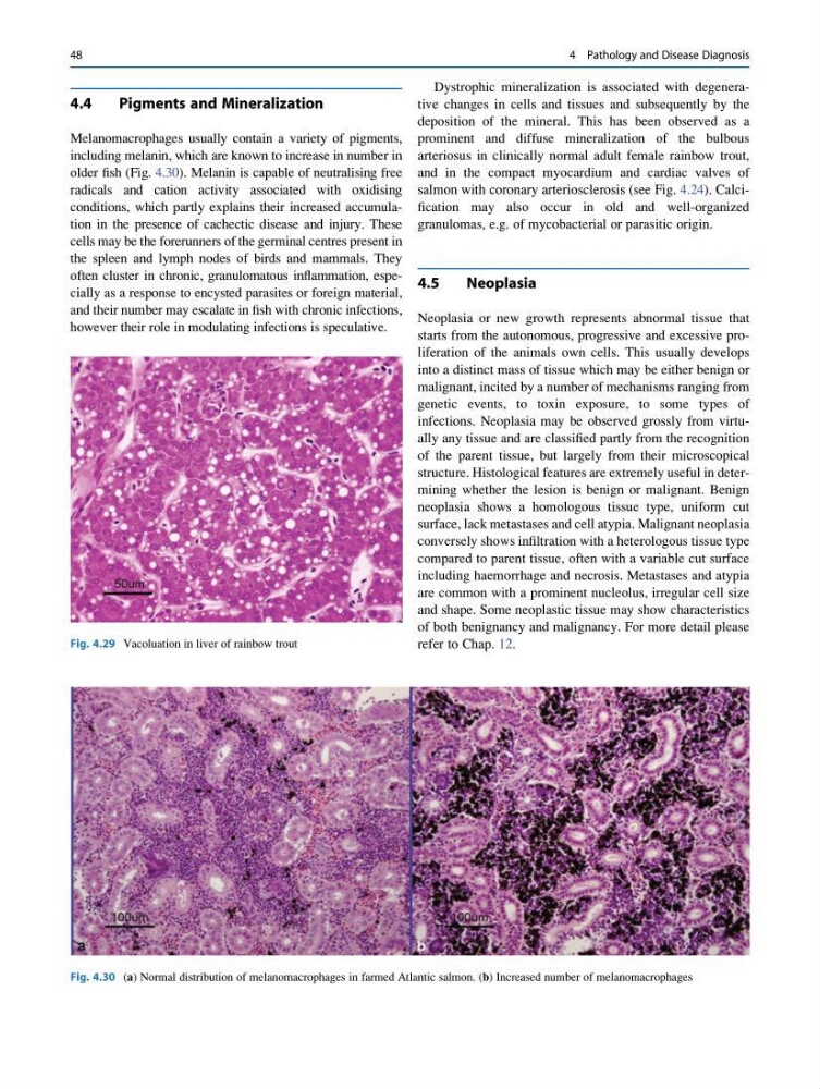
Melanomacrophages usually contain a variety of pigments,
including melanin, which are known to increase in number in
older fish (Fig. 4.30). Melanin is capable of neutralising free
radicals and cation activity associated with oxidising
conditions, which partly explains their increased accumulation in the presence of cachectic disease and injury. These
cells may be the forerunners of the germinal centres present in
the spleen and lymph nodes of birds and mammals. They
often cluster in chronic, granulomatous inflammation, especially as a response to encysted parasites or foreign material,
and their number may escalate in fish with chronic infections,
however their role in modulating infections is speculative.
Dystrophic mineralization is associated with degenerative changes in cells and tissues and subsequently by the
deposition of the mineral. This has been observed as a
prominent and diffuse mineralization of the bulbous
arteriosus in clinically normal adult female rainbow trout,
and in the compact myocardium and cardiac valves of
salmon with coronary arteriosclerosis (see Fig. 4.24). Calcification may also occur in old and well-organized
granulomas, e.g. of mycobacterial or parasitic origin.
4.5
Neoplasia
Neoplasia or new growth represents abnormal tissue that
starts from the autonomous, progressive and excessive proliferation of the animals own cells. This usually develops
into a distinct mass of tissue which may be either benign or
malignant, incited by a number of mechanisms ranging from
genetic events, to toxin exposure, to some types of
infections. Neoplasia may be observed grossly from virtually any tissue and are classified partly from the recognition
of the parent tissue, but largely from their microscopical
structure. Histological features are extremely useful in determining whether the lesion is benign or malignant. Benign
neoplasia shows a homologous tissue type, uniform cut
surface, lack metastases and cell atypia. Malignant neoplasia
conversely shows infiltration with a heterologous tissue type
compared to parent tissue, often with a variable cut surface
including haemorrhage and necrosis. Metastases and atypia
are common with a prominent nucleolus, irregular cell size
and shape. Some neoplastic tissue may show characteristics
of both benignancy and malignancy. For more detail please
refer to Chap. 12.
Fig. 4.30 (a) Normal distribution of melanomacrophages in farmed Atlantic salmon. (b) Increased number of melanomacrophages
Fig. 4.29 Vacoluation in liver of rainbow trout
48
4 Pathology and Disease Diagnosis
Pigments and Mineralization
Melanomacrophages usually contain a variety of pigments,
including melanin, which are known to increase in number in
older fish (Fig. 4.30). Melanin is capable of neutralising free
radicals and cation activity associated with oxidising
conditions, which partly explains their increased accumulation in the presence of cachectic disease and injury. These
cells may be the forerunners of the germinal centres present in
the spleen and lymph nodes of birds and mammals. They
often cluster in chronic, granulomatous inflammation, especially as a response to encysted parasites or foreign material,
and their number may escalate in fish with chronic infections,
however their role in modulating infections is speculative.
Dystrophic mineralization is associated with degenerative changes in cells and tissues and subsequently by the
deposition of the mineral. This has been observed as a
prominent and diffuse mineralization of the bulbous
arteriosus in clinically normal adult female rainbow trout,
and in the compact myocardium and cardiac valves of
salmon with coronary arteriosclerosis (see Fig. 4.24). Calcification may also occur in old and well-organized
granulomas, e.g. of mycobacterial or parasitic origin.
4.5
Neoplasia
Neoplasia or new growth represents abnormal tissue that
starts from the autonomous, progressive and excessive proliferation of the animals own cells. This usually develops
into a distinct mass of tissue which may be either benign or
malignant, incited by a number of mechanisms ranging from
genetic events, to toxin exposure, to some types of
infections. Neoplasia may be observed grossly from virtually any tissue and are classified partly from the recognition
of the parent tissue, but largely from their microscopical
structure. Histological features are extremely useful in determining whether the lesion is benign or malignant. Benign
neoplasia shows a homologous tissue type, uniform cut
surface, lack metastases and cell atypia. Malignant neoplasia
conversely shows infiltration with a heterologous tissue type
compared to parent tissue, often with a variable cut surface
including haemorrhage and necrosis. Metastases and atypia
are common with a prominent nucleolus, irregular cell size
and shape. Some neoplastic tissue may show characteristics
of both benignancy and malignancy. For more detail please
refer to Chap. 12.
Fig. 4.30 (a) Normal distribution of melanomacrophages in farmed Atlantic salmon. (b) Increased number of melanomacrophages
Fig. 4.29 Vacoluation in liver of rainbow trout
48
4 Pathology and Disease Diagnosis