

beneath consisting of non-nucleated long, slender, transparent cells in parallel rows that occupy the greatest volume.
The lens protrudes through the iris providing a wide angle of
view. In order to accommodate changing sight requirements,
the retractor lentis muscle must be drawn inwards towards
the retina. The iris is fixed as the sphincter and the dilator
muscles are poorly developed. The innermost element of the
eyeball is the photo-sensitive retina. Choroid vessels form a
subscleral network of capillaries which provide nourishment
for the retina. Eight layers are recognized: pigment epithelium elements, rods and cones, outer nuclear layer, outer
plexiform layer, inner nuclear layer, inner plexiform layer,
ganglion cell layer and nerve fibre layer (Fig. 2.42).
A counter current system is present in the choroid layer of
the retina. This vascular structure (the choroid rete mirabile)
is supplied by a branch of the ophthalmic artery and blood
from of the choroidal vessels initially passes through the
pseudobranch. The correlation between the oxygen pressure
and development of the rete suggest that the choroid rete
mirabile plays a role in establishing the high oxygen pressure of the retina.
2.8
Nervous System
The nervous system can be divided into the cerebrospinal
system, i.e., the brain, spinal cord, ganglia, cranial and spinal
nerves, and the autonomous or vegetative system, comprising ganglia and sympathetic and parasympathetic nerves that
works in close integration and interdependency with the
endocrine system. Overall the main function is integration
and control of organs and the communication with the outside environment. Histologically, the nervous system cellular elements are the neurons and the neuroglia and consist of
two basic types of cell: neurons and glial cells.
2.8.1 Brain
The brain has the same basic regions as that of other
vertebrates, but the proportions between the anatomical
units are different, particularly the mesencephalon with the
optic lobes being very conspicuous (Fig. 2.43). The brain is
traditionally divided into five different units: telencephalon,
diencephalon, mesencephalon, metencephalon and myelencephalon. The protective layers around the brain are known
as the meninges, and represent an important barrier against
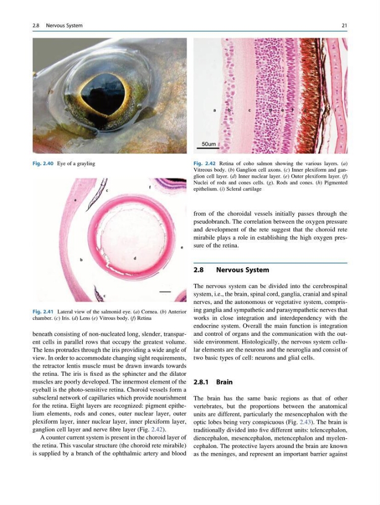
Fig. 2.40 Eye of a grayling
Fig. 2.41 Lateral view of the salmonid eye. (a) Cornea. (b) Anterior
chamber. (c) Iris. (d) Lens (e) Vitrous body. (f) Retina
Fig. 2.42 Retina of coho salmon showing the various layers. (a)
Vitreous body. (b) Ganglion cell axons. (c) Inner plexiform and ganglion cell layer. (d) Inner nuclear layer. (e) Outer plexiform layer. (f)
Nuclei of rods and cones cells. (g). Rods and cones. (h) Pigmented
epithelium. (i) Scleral cartilage
2.8 Nervous System
21
The lens protrudes through the iris providing a wide angle of
view. In order to accommodate changing sight requirements,
the retractor lentis muscle must be drawn inwards towards
the retina. The iris is fixed as the sphincter and the dilator
muscles are poorly developed. The innermost element of the
eyeball is the photo-sensitive retina. Choroid vessels form a
subscleral network of capillaries which provide nourishment
for the retina. Eight layers are recognized: pigment epithelium elements, rods and cones, outer nuclear layer, outer
plexiform layer, inner nuclear layer, inner plexiform layer,
ganglion cell layer and nerve fibre layer (Fig. 2.42).
A counter current system is present in the choroid layer of
the retina. This vascular structure (the choroid rete mirabile)
is supplied by a branch of the ophthalmic artery and blood
from of the choroidal vessels initially passes through the
pseudobranch. The correlation between the oxygen pressure
and development of the rete suggest that the choroid rete
mirabile plays a role in establishing the high oxygen pressure of the retina.
2.8
Nervous System
The nervous system can be divided into the cerebrospinal
system, i.e., the brain, spinal cord, ganglia, cranial and spinal
nerves, and the autonomous or vegetative system, comprising ganglia and sympathetic and parasympathetic nerves that
works in close integration and interdependency with the
endocrine system. Overall the main function is integration
and control of organs and the communication with the outside environment. Histologically, the nervous system cellular elements are the neurons and the neuroglia and consist of
two basic types of cell: neurons and glial cells.
2.8.1 Brain
The brain has the same basic regions as that of other
vertebrates, but the proportions between the anatomical
units are different, particularly the mesencephalon with the
optic lobes being very conspicuous (Fig. 2.43). The brain is
traditionally divided into five different units: telencephalon,
diencephalon, mesencephalon, metencephalon and myelencephalon. The protective layers around the brain are known
as the meninges, and represent an important barrier against
Fig. 2.40 Eye of a grayling
Fig. 2.41 Lateral view of the salmonid eye. (a) Cornea. (b) Anterior
chamber. (c) Iris. (d) Lens (e) Vitrous body. (f) Retina
Fig. 2.42 Retina of coho salmon showing the various layers. (a)
Vitreous body. (b) Ganglion cell axons. (c) Inner plexiform and ganglion cell layer. (d) Inner nuclear layer. (e) Outer plexiform layer. (f)
Nuclei of rods and cones cells. (g). Rods and cones. (h) Pigmented
epithelium. (i) Scleral cartilage
2.8 Nervous System
21