

2.7
Sensory System
Fish sensory systems include mechano, olfactory and taste
receptors, equilibrium, hearing as well as vision organs. The
olfactory sensory neurons give a sense of smell mediated by
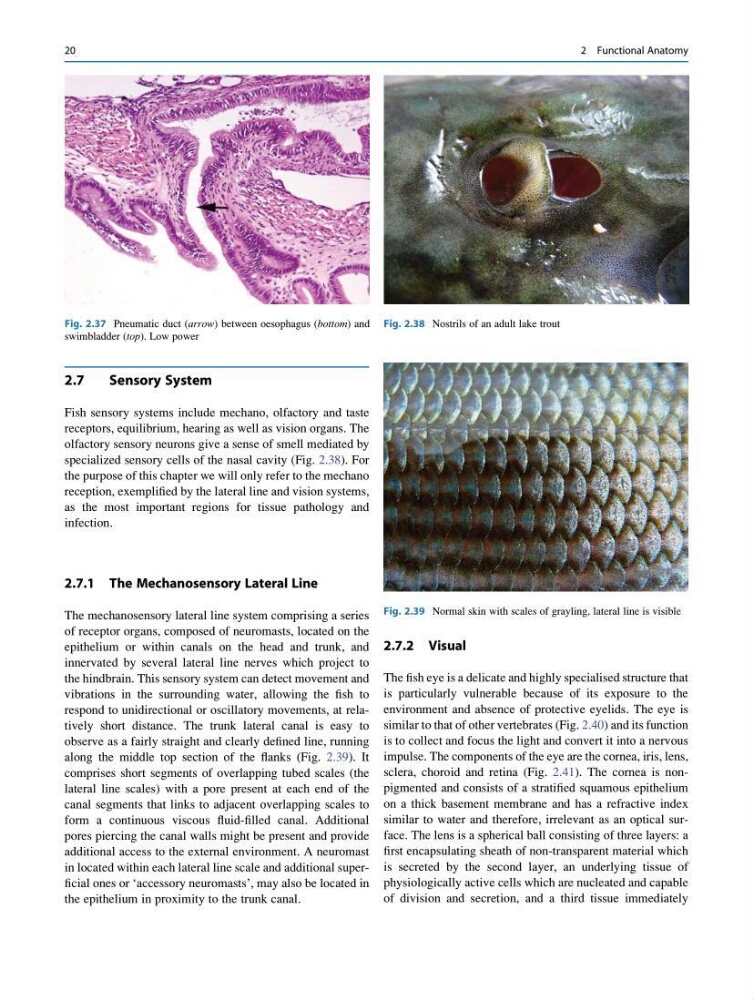
specialized sensory cells of the nasal cavity (Fig. 2.38). For
the purpose of this chapter we will only refer to the mechano
reception, exemplified by the lateral line and vision systems,
as the most important regions for tissue pathology and
infection.
2.7.1 The Mechanosensory Lateral Line
The mechanosensory lateral line system comprising a series
of receptor organs, composed of neuromasts, located on the
epithelium or within canals on the head and trunk, and
innervated by several lateral line nerves which project to
the hindbrain. This sensory system can detect movement and
vibrations in the surrounding water, allowing the fish to
respond to unidirectional or oscillatory movements, at relatively short distance. The trunk lateral canal is easy to
observe as a fairly straight and clearly defined line, running
along the middle top section of the flanks (Fig. 2.39). It
comprises short segments of overlapping tubed scales (the
lateral line scales) with a pore present at each end of the
canal segments that links to adjacent overlapping scales to
form a continuous viscous fluid-filled canal. Additional
pores piercing the canal walls might be present and provide
additional access to the external environment. A neuromast
in located within each lateral line scale and additional superficial ones or ‘accessory neuromasts’, may also be located in
the epithelium in proximity to the trunk canal.
2.7.2 Visual
The fish eye is a delicate and highly specialised structure that
is particularly vulnerable because of its exposure to the
environment and absence of protective eyelids. The eye is
similar to that of other vertebrates (Fig. 2.40) and its function
is to collect and focus the light and convert it into a nervous
impulse. The components of the eye are the cornea, iris, lens,
sclera, choroid and retina (Fig. 2.41). The cornea is nonpigmented and consists of a stratified squamous epithelium
on a thick basement membrane and has a refractive index
similar to water and therefore, irrelevant as an optical surface. The lens is a spherical ball consisting of three layers: a
first encapsulating sheath of non-transparent material which
is secreted by the second layer, an underlying tissue of
physiologically active cells which are nucleated and capable
of division and secretion, and a third tissue immediately
Fig. 2.37 Pneumatic duct (arrow) between oesophagus (bottom) and
swimbladder (top). Low power
Fig. 2.38 Nostrils of an adult lake trout
Fig. 2.39 Normal skin with scales of grayling, lateral line is visible
20
2 Functional Anatomy
Sensory System
Fish sensory systems include mechano, olfactory and taste
receptors, equilibrium, hearing as well as vision organs. The
olfactory sensory neurons give a sense of smell mediated by
specialized sensory cells of the nasal cavity (Fig. 2.38). For
the purpose of this chapter we will only refer to the mechano
reception, exemplified by the lateral line and vision systems,
as the most important regions for tissue pathology and
infection.
2.7.1 The Mechanosensory Lateral Line
The mechanosensory lateral line system comprising a series
of receptor organs, composed of neuromasts, located on the
epithelium or within canals on the head and trunk, and
innervated by several lateral line nerves which project to
the hindbrain. This sensory system can detect movement and
vibrations in the surrounding water, allowing the fish to
respond to unidirectional or oscillatory movements, at relatively short distance. The trunk lateral canal is easy to
observe as a fairly straight and clearly defined line, running
along the middle top section of the flanks (Fig. 2.39). It
comprises short segments of overlapping tubed scales (the
lateral line scales) with a pore present at each end of the
canal segments that links to adjacent overlapping scales to
form a continuous viscous fluid-filled canal. Additional
pores piercing the canal walls might be present and provide
additional access to the external environment. A neuromast
in located within each lateral line scale and additional superficial ones or ‘accessory neuromasts’, may also be located in
the epithelium in proximity to the trunk canal.
2.7.2 Visual
The fish eye is a delicate and highly specialised structure that
is particularly vulnerable because of its exposure to the
environment and absence of protective eyelids. The eye is
similar to that of other vertebrates (Fig. 2.40) and its function
is to collect and focus the light and convert it into a nervous
impulse. The components of the eye are the cornea, iris, lens,
sclera, choroid and retina (Fig. 2.41). The cornea is nonpigmented and consists of a stratified squamous epithelium
on a thick basement membrane and has a refractive index
similar to water and therefore, irrelevant as an optical surface. The lens is a spherical ball consisting of three layers: a
first encapsulating sheath of non-transparent material which
is secreted by the second layer, an underlying tissue of
physiologically active cells which are nucleated and capable
of division and secretion, and a third tissue immediately
Fig. 2.37 Pneumatic duct (arrow) between oesophagus (bottom) and
swimbladder (top). Low power
Fig. 2.38 Nostrils of an adult lake trout
Fig. 2.39 Normal skin with scales of grayling, lateral line is visible
20
2 Functional Anatomy