

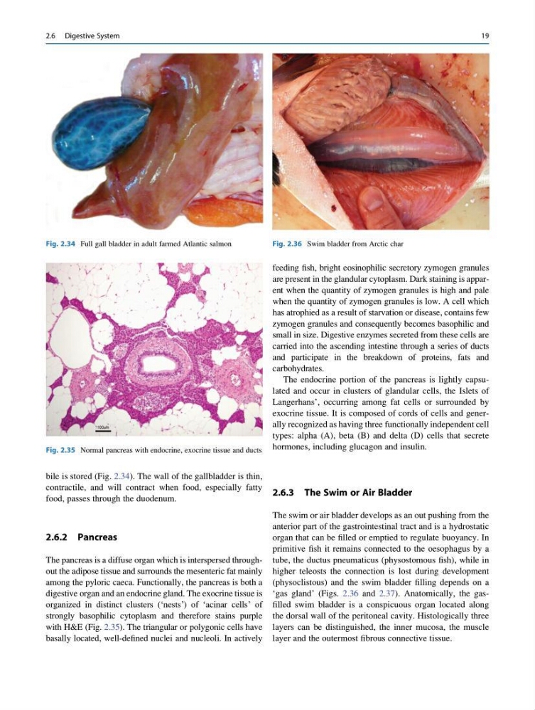
bile is stored (Fig. 2.34). The wall of the gallbladder is thin,
contractile, and will contract when food, especially fatty
food, passes through the duodenum.
2.6.2 Pancreas
The pancreas is a diffuse organ which is interspersed throughout the adipose tissue and surrounds the mesenteric fat mainly
among the pyloric caeca. Functionally, the pancreas is both a
digestive organ and an endocrine gland. The exocrine tissue is
organized in distinct clusters (‘nests’) of ‘acinar cells’ of
strongly basophilic cytoplasm and therefore stains purple
with H&E (Fig. 2.35). The triangular or polygonic cells have
basally located, well-defined nuclei and nucleoli. In actively
feeding fish, bright eosinophilic secretory zymogen granules
are present in the glandular cytoplasm. Dark staining is apparent when the quantity of zymogen granules is high and pale
when the quantity of zymogen granules is low. A cell which
has atrophied as a result of starvation or disease, contains few
zymogen granules and consequently becomes basophilic and
small in size. Digestive enzymes secreted from these cells are
carried into the ascending intestine through a series of ducts
and participate in the breakdown of proteins, fats and
carbohydrates.
The endocrine portion of the pancreas is lightly capsulated and occur in clusters of glandular cells, the Islets of
Langerhans’, occurring among fat cells or surrounded by
exocrine tissue. It is composed of cords of cells and generally recognized as having three functionally independent cell
types: alpha (A), beta (B) and delta (D) cells that secrete
hormones, including glucagon and insulin.
2.6.3 The Swim or Air Bladder
The swim or air bladder develops as an out pushing from the
anterior part of the gastrointestinal tract and is a hydrostatic
organ that can be filled or emptied to regulate buoyancy. In
primitive fish it remains connected to the oesophagus by a
tube, the ductus pneumaticus (physostomous fish), while in
higher teleosts the connection is lost during development
(physoclistous) and the swim bladder filling depends on a
‘gas gland’ (Figs. 2.36 and 2.37). Anatomically, the gasfilled swim bladder is a conspicuous organ located along
the dorsal wall of the peritoneal cavity. Histologically three
layers can be distinguished, the inner mucosa, the muscle
layer and the outermost fibrous connective tissue.
Fig. 2.34 Full gall bladder in adult farmed Atlantic salmon
Fig. 2.35 Normal pancreas with endocrine, exocrine tissue and ducts
Fig. 2.36 Swim bladder from Arctic char
2.6 Digestive System
19
contractile, and will contract when food, especially fatty
food, passes through the duodenum.
2.6.2 Pancreas
The pancreas is a diffuse organ which is interspersed throughout the adipose tissue and surrounds the mesenteric fat mainly
among the pyloric caeca. Functionally, the pancreas is both a
digestive organ and an endocrine gland. The exocrine tissue is
organized in distinct clusters (‘nests’) of ‘acinar cells’ of
strongly basophilic cytoplasm and therefore stains purple
with H&E (Fig. 2.35). The triangular or polygonic cells have
basally located, well-defined nuclei and nucleoli. In actively
feeding fish, bright eosinophilic secretory zymogen granules
are present in the glandular cytoplasm. Dark staining is apparent when the quantity of zymogen granules is high and pale
when the quantity of zymogen granules is low. A cell which
has atrophied as a result of starvation or disease, contains few
zymogen granules and consequently becomes basophilic and
small in size. Digestive enzymes secreted from these cells are
carried into the ascending intestine through a series of ducts
and participate in the breakdown of proteins, fats and
carbohydrates.
The endocrine portion of the pancreas is lightly capsulated and occur in clusters of glandular cells, the Islets of
Langerhans’, occurring among fat cells or surrounded by
exocrine tissue. It is composed of cords of cells and generally recognized as having three functionally independent cell
types: alpha (A), beta (B) and delta (D) cells that secrete
hormones, including glucagon and insulin.
2.6.3 The Swim or Air Bladder
The swim or air bladder develops as an out pushing from the
anterior part of the gastrointestinal tract and is a hydrostatic
organ that can be filled or emptied to regulate buoyancy. In
primitive fish it remains connected to the oesophagus by a
tube, the ductus pneumaticus (physostomous fish), while in
higher teleosts the connection is lost during development
(physoclistous) and the swim bladder filling depends on a
‘gas gland’ (Figs. 2.36 and 2.37). Anatomically, the gasfilled swim bladder is a conspicuous organ located along
the dorsal wall of the peritoneal cavity. Histologically three
layers can be distinguished, the inner mucosa, the muscle
layer and the outermost fibrous connective tissue.
Fig. 2.34 Full gall bladder in adult farmed Atlantic salmon
Fig. 2.35 Normal pancreas with endocrine, exocrine tissue and ducts
Fig. 2.36 Swim bladder from Arctic char
2.6 Digestive System
19