

folds. In rainbow trout for example, the anterior region has
shallow folds and the posterior region deep folds with a
thicker muscle layer with many mucous cells.
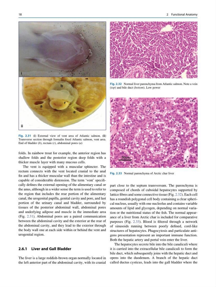
The vent is equipped with a muscular sphincter. The
rectum connects with the vent located cranial to the anal
fin and has a thicker muscular wall than the intestine and is
capable of considerable distension. The term ‘vent’ specifically defines the external opening of the alimentary canal or
the anus, although in a wider sense the term is used to refer to
the region that includes the rear portion of the alimentary
canal, the urogenital papilla, genital cavity and pore, and last
portion of the urinary canal and bladder, surrounded by
tissues of the posterior abdominal wall, abdominal pores
and underlying adipose and muscle in the immediate area
(Fig. 2.31). Abdominal pores are a paired communication
between the abdominal cavity and the exterior at the rear of
the abdominal cavity, and they lead to the exterior through
the body wall one at each side within or behind the vent and
urogenital region.
2.6.1 Liver and Gall Bladder
The liver is a large reddish-brown organ normally located in
the left anterior part of the abdominal cavity, with its cranial
part close to the septum transversum. The parenchyma is
composed of chords of cuboidal hepatocytes supported by
lattice fibres and some connective tissue (Fig. 2.32). Each cell
has a roundish polygonal cell body containing a clear spherical nucleus, usually with one nucleolus and contains variable
amounts of lipid and glycogen, depending on normal variation or the nutritional status of the fish. The normal appearance of a liver from Arctic char is included for comparative
purposes (Fig. 2.33). Blood is filtered through a network
of sinusoids running between poorly defined, cord-like
structures of hepatocytes. Phagocytosis and particulate antigens presentation represent an important immune function.
Both the hepatic artery and portal vein enter the liver.
The hepatocytes secrete bile into the bile canaliculi where
it is carried into the extracellular bile canaliculi to form the
bile duct, which subsequently joins with the hepatic duct and
opens into the duodenum. A branch of the hepatic duct
called ductus cysticus, leads into the gall bladder where the
Fig. 2.31 (i) External view of vent area of Atlantic salmon. (ii)
Transverse section through formalin fixed Atlantic salmon, vent area.
End of bladder (b), rectum (r), abdominal pores (a)
Fig. 2.32 Normal liver parenchyma from Atlantic salmon. Note a vein
(top) and bile duct (bottom). Low power
Fig. 2.33 Normal parenchyma of Arctic char liver
18
2 Functional Anatomy
shallow folds and the posterior region deep folds with a
thicker muscle layer with many mucous cells.
The vent is equipped with a muscular sphincter. The
rectum connects with the vent located cranial to the anal
fin and has a thicker muscular wall than the intestine and is
capable of considerable distension. The term ‘vent’ specifically defines the external opening of the alimentary canal or
the anus, although in a wider sense the term is used to refer to
the region that includes the rear portion of the alimentary
canal, the urogenital papilla, genital cavity and pore, and last
portion of the urinary canal and bladder, surrounded by
tissues of the posterior abdominal wall, abdominal pores
and underlying adipose and muscle in the immediate area
(Fig. 2.31). Abdominal pores are a paired communication
between the abdominal cavity and the exterior at the rear of
the abdominal cavity, and they lead to the exterior through
the body wall one at each side within or behind the vent and
urogenital region.
2.6.1 Liver and Gall Bladder
The liver is a large reddish-brown organ normally located in
the left anterior part of the abdominal cavity, with its cranial
part close to the septum transversum. The parenchyma is
composed of chords of cuboidal hepatocytes supported by
lattice fibres and some connective tissue (Fig. 2.32). Each cell
has a roundish polygonal cell body containing a clear spherical nucleus, usually with one nucleolus and contains variable
amounts of lipid and glycogen, depending on normal variation or the nutritional status of the fish. The normal appearance of a liver from Arctic char is included for comparative
purposes (Fig. 2.33). Blood is filtered through a network
of sinusoids running between poorly defined, cord-like
structures of hepatocytes. Phagocytosis and particulate antigens presentation represent an important immune function.
Both the hepatic artery and portal vein enter the liver.
The hepatocytes secrete bile into the bile canaliculi where
it is carried into the extracellular bile canaliculi to form the
bile duct, which subsequently joins with the hepatic duct and
opens into the duodenum. A branch of the hepatic duct
called ductus cysticus, leads into the gall bladder where the
Fig. 2.31 (i) External view of vent area of Atlantic salmon. (ii)
Transverse section through formalin fixed Atlantic salmon, vent area.
End of bladder (b), rectum (r), abdominal pores (a)
Fig. 2.32 Normal liver parenchyma from Atlantic salmon. Note a vein
(top) and bile duct (bottom). Low power
Fig. 2.33 Normal parenchyma of Arctic char liver
18
2 Functional Anatomy