

folds (crypts). The pharynx mucosa contains a lamina
propria, but is devoid of a muscularis mucosa. The
muscularis is composed of a thick outer layer of circular
muscle and a thin layer of longitudinal muscle. Both layers
of the muscularis are striated.
The oesophagus is a short, muscular thick-walled tube
with longitudinally arranged folds of mucosa to facilitate
swallowing and propulsion of food particles. At the entrance
to the oesophagus the mucosa contains many mucous cells
and taste buds. This region usually lacks a muscularis
mucosa, distinguishing it from the rest of the digestive tract.
In the stomach and intestine the stratum compactum is
located between the lamina propria and the muscularis
mucosa which is composed of dense collagen fibres. Eosinophilic granular cells (EGC’s) may be present and form a layer
at the inner and outer sides of this stratum and are considered
to be closely related to fish mast cells. This layer of EGC’s is
sometimes referred to as the stratum granulosum.
The stomach is U-shaped and composed of the cardiac
portion (anterior), fundus and pyloric portion. Each region
has a simple folded mucosal epithelium. At the cardiac portion the folds are shallow and become deeper at the fundus
and pyloric portion. The epithelium varies between cuboidal
and highly columnar, and the nuclei are generally located in
the basal region of the cell. Gastric glands are located in the
lamina propria and often into the crypts of the mucosal folds.
The fundic region of the stomach is a blind sac, pouching
off from the main tube of the organ and characterized by
numerous gastric glands although, in contrast, they may be
absent in the pyloric region.
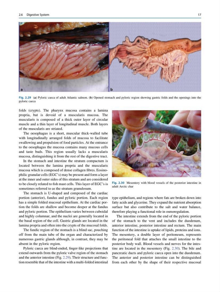
Pyloric caeca are blind-ended, finger-like projections that
extend outwards from the pyloric valve region of the stomach
and the anterior intestine (Fig. 2.29). Their structure and function resemble that of the intestine with a multi-folded intestinal
type epithelium, and regions where fats are broken down into
fatty acids and glycerine. They expand the nutrient absorption
surface but also contribute to the salt and water balance,
therefore playing a functional role in osmoregulation.
The intestine extends from the end of the pyloric portion
of the stomach to the vent and includes the duodenum,
anterior intestine, posterior intestine and rectum. The main
function of the intestine is uptake of lipids, proteins and ions.
The mesentery, a double layer of peritoneum, represents
the peritoneal fold that attaches the small intestine to the
posterior body wall. Blood vessels and nerves for the intestine are located in the mesentery (Fig. 2.30). The bile and
pancreatic ducts and pyloric caeca open into the duodenum.
The anterior and posterior intestine can be distinguished
from each other by the shape of their respective mucosal
Fig. 2.29 (a) Pyloric caeca of adult Atlantic salmon. (b) Opened stomach and pyloric region showing gastric folds and the openings into the
pyloric caeca
Fig. 2.30 Mesentery with blood vessels of the posterior intestine in
adult Arctic char
2.6 Digestive System
17
propria, but is devoid of a muscularis mucosa. The
muscularis is composed of a thick outer layer of circular
muscle and a thin layer of longitudinal muscle. Both layers
of the muscularis are striated.
The oesophagus is a short, muscular thick-walled tube
with longitudinally arranged folds of mucosa to facilitate
swallowing and propulsion of food particles. At the entrance
to the oesophagus the mucosa contains many mucous cells
and taste buds. This region usually lacks a muscularis
mucosa, distinguishing it from the rest of the digestive tract.
In the stomach and intestine the stratum compactum is
located between the lamina propria and the muscularis
mucosa which is composed of dense collagen fibres. Eosinophilic granular cells (EGC’s) may be present and form a layer
at the inner and outer sides of this stratum and are considered
to be closely related to fish mast cells. This layer of EGC’s is
sometimes referred to as the stratum granulosum.
The stomach is U-shaped and composed of the cardiac
portion (anterior), fundus and pyloric portion. Each region
has a simple folded mucosal epithelium. At the cardiac portion the folds are shallow and become deeper at the fundus
and pyloric portion. The epithelium varies between cuboidal
and highly columnar, and the nuclei are generally located in
the basal region of the cell. Gastric glands are located in the
lamina propria and often into the crypts of the mucosal folds.
The fundic region of the stomach is a blind sac, pouching
off from the main tube of the organ and characterized by
numerous gastric glands although, in contrast, they may be
absent in the pyloric region.
Pyloric caeca are blind-ended, finger-like projections that
extend outwards from the pyloric valve region of the stomach
and the anterior intestine (Fig. 2.29). Their structure and function resemble that of the intestine with a multi-folded intestinal
type epithelium, and regions where fats are broken down into
fatty acids and glycerine. They expand the nutrient absorption
surface but also contribute to the salt and water balance,
therefore playing a functional role in osmoregulation.
The intestine extends from the end of the pyloric portion
of the stomach to the vent and includes the duodenum,
anterior intestine, posterior intestine and rectum. The main
function of the intestine is uptake of lipids, proteins and ions.
The mesentery, a double layer of peritoneum, represents
the peritoneal fold that attaches the small intestine to the
posterior body wall. Blood vessels and nerves for the intestine are located in the mesentery (Fig. 2.30). The bile and
pancreatic ducts and pyloric caeca open into the duodenum.
The anterior and posterior intestine can be distinguished
from each other by the shape of their respective mucosal
Fig. 2.29 (a) Pyloric caeca of adult Atlantic salmon. (b) Opened stomach and pyloric region showing gastric folds and the openings into the
pyloric caeca
Fig. 2.30 Mesentery with blood vessels of the posterior intestine in
adult Arctic char
2.6 Digestive System
17