

occupies the centre of the tooth. Odontoblasts are arranged
at the outermost region of the pulp and secrete dentin.
The presence of teeth does not imply chewing activity but
often have a role in grabbing and tearing food.
Although the histological structure of the alimentary
canal varies along its length, it is basically composed of
the following layers from inside out: mucosa, submucosa,
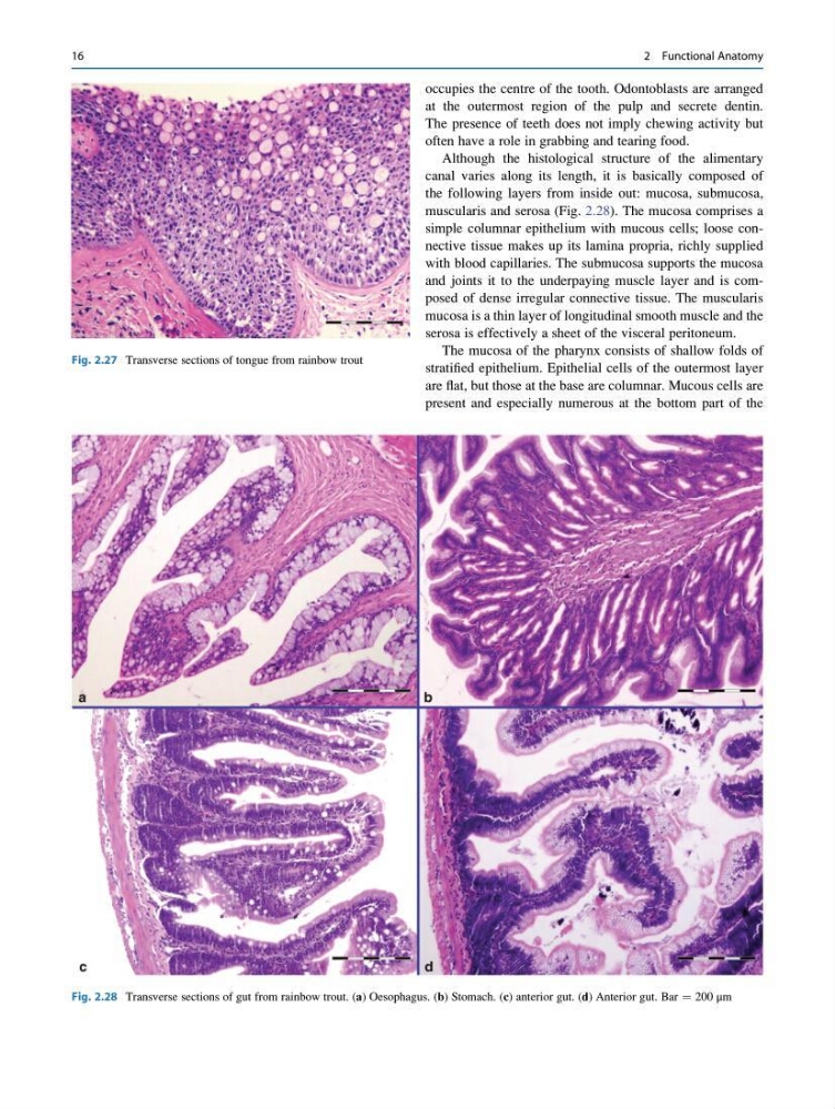
muscularis and serosa (Fig. 2.28). The mucosa comprises a
simple columnar epithelium with mucous cells; loose connective tissue makes up its lamina propria, richly supplied
with blood capillaries. The submucosa supports the mucosa
and joints it to the underpaying muscle layer and is composed of dense irregular connective tissue. The muscularis
mucosa is a thin layer of longitudinal smooth muscle and the
serosa is effectively a sheet of the visceral peritoneum.
The mucosa of the pharynx consists of shallow folds of
stratified epithelium. Epithelial cells of the outermost layer
are flat, but those at the base are columnar. Mucous cells are
present and especially numerous at the bottom part of the
Fig. 2.27 Transverse sections of tongue from rainbow trout
Fig. 2.28 Transverse sections of gut from rainbow trout. (a) Oesophagus. (b) Stomach. (c) anterior gut. (d) Anterior gut. Bar ¼ 200 μm
16
2 Functional Anatomy
at the outermost region of the pulp and secrete dentin.
The presence of teeth does not imply chewing activity but
often have a role in grabbing and tearing food.
Although the histological structure of the alimentary
canal varies along its length, it is basically composed of
the following layers from inside out: mucosa, submucosa,
muscularis and serosa (Fig. 2.28). The mucosa comprises a
simple columnar epithelium with mucous cells; loose connective tissue makes up its lamina propria, richly supplied
with blood capillaries. The submucosa supports the mucosa
and joints it to the underpaying muscle layer and is composed of dense irregular connective tissue. The muscularis
mucosa is a thin layer of longitudinal smooth muscle and the
serosa is effectively a sheet of the visceral peritoneum.
The mucosa of the pharynx consists of shallow folds of
stratified epithelium. Epithelial cells of the outermost layer
are flat, but those at the base are columnar. Mucous cells are
present and especially numerous at the bottom part of the
Fig. 2.27 Transverse sections of tongue from rainbow trout
Fig. 2.28 Transverse sections of gut from rainbow trout. (a) Oesophagus. (b) Stomach. (c) anterior gut. (d) Anterior gut. Bar ¼ 200 μm
16
2 Functional Anatomy