

pathogens between the blood and the cerebrospinal fluid
(Fig. 2.44). They are small compared to its mammalian
counterparts and are dominated by the olfactory lobes. In
contrast to mammals, the histological conspicuous sixlayered neocortex is absent.
The ventral part of the diencephalon, including the
hypothalamus, is termed the infundibulum and is well
developed in salmonids. Three important proteins are produced here and important components of the cerebrospinal
fluid.
The telencephalon structures are located in the ventral
midline of the diencephalon: chiasmaopticus, the pituitary
gland (hypophysis) and a ventral choroid plexus called
‘saccus vasculosus’. In the optic chiasma, the nerve fibres
from the retina are crossing before entering the brain. The
complex pituitary gland is a neuro-epithelial structure
orchestrating several other endocrine organs and is
involved in osmoregulation, gonadal development, growth
and melanization. The saccus vasculosus is a sac-like structure with blood sinusoids quite similar to the choroid
plexus of the eye, and communicates with the lumen of
the third ventricle. On the dorsal side of the diencephalon is
the pineal gland (epiphysis). This is a well vascularised
non-image forming photoreceptor structure, located under
a thin non-pigmented area of the skull that permits light to
reach the structure (‘pineal window’). This structure can
detect alterations in ambient light (decreasing light in fall,
increasing light in the spring) and is of importance for
regulation of seasonal physiology such as smoltification.
The mesencephalon is highly developed and conspicuous
with its two optic lobes (tectumopticum) reflecting the
importance of vision for these fish. Centrally, there is a
large lumen (ventriculus mesencephali) filled with cerebrospinal fluid. The metencephalon has a well-developed,
partly folded dorsal part called the cerebellum, responsible
for movement coordination. The myelencephalon is the
origin of the cranial nerves and the beginning of the spinal
cord (medulla oblongata).
2.8.2 The Sympathetic and Parasympathetic
Ganglions and Nerves
The sympathetic and parasympathetic ganglions and nerves
of the autonomous nervous system are responsible of
regulating several functions and mostly antagonist
responses to that of the cerebrospinal system, as both
innervate most internal organs including digestive tract,
heart and gills.
2.9
Endocrine System
Glandular derivatives such as thyroid, thymus and
ultimobranchial bodies are formed from the pharynx during
embryological development. Thyroid hormones play a supportive role in sea water acclimation and follicles are
distributed throughout the connective tissue of the pharyngeal area. They may also be observed around the eye, ventral
aorta, hepatic veins and anterior kidney, and are similar
histologically to mammalian thyroid tissue (Fig. 2.45). The
thymus is located on the dorsolateral wall of the pharynx
with its ventral surface covered with mucosal epithelium
(Fig. 2.46).
At the junction of the head and posterior kidney are the
corpuscles of Stannius, a sac-like body which has an endocrine role in calcium metabolism. The corpuscles of
Stannius are not always visible macroscopically and may
be embedded deep in the renal tissue (Figs. 2.47 and 2.48).
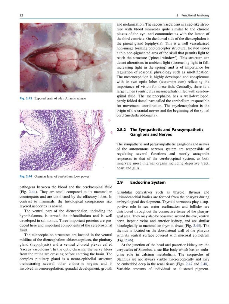
Variable amounts of individual or clustered pigmentFig. 2.43 Exposed brain of adult Atlantic salmon
Fig. 2.44 Granular layer of cerebellum. Low power
22
2 Functional Anatomy
(Fig. 2.44). They are small compared to its mammalian
counterparts and are dominated by the olfactory lobes. In
contrast to mammals, the histological conspicuous sixlayered neocortex is absent.
The ventral part of the diencephalon, including the
hypothalamus, is termed the infundibulum and is well
developed in salmonids. Three important proteins are produced here and important components of the cerebrospinal
fluid.
The telencephalon structures are located in the ventral
midline of the diencephalon: chiasmaopticus, the pituitary
gland (hypophysis) and a ventral choroid plexus called
‘saccus vasculosus’. In the optic chiasma, the nerve fibres
from the retina are crossing before entering the brain. The
complex pituitary gland is a neuro-epithelial structure
orchestrating several other endocrine organs and is
involved in osmoregulation, gonadal development, growth
and melanization. The saccus vasculosus is a sac-like structure with blood sinusoids quite similar to the choroid
plexus of the eye, and communicates with the lumen of
the third ventricle. On the dorsal side of the diencephalon is
the pineal gland (epiphysis). This is a well vascularised
non-image forming photoreceptor structure, located under
a thin non-pigmented area of the skull that permits light to
reach the structure (‘pineal window’). This structure can
detect alterations in ambient light (decreasing light in fall,
increasing light in the spring) and is of importance for
regulation of seasonal physiology such as smoltification.
The mesencephalon is highly developed and conspicuous
with its two optic lobes (tectumopticum) reflecting the
importance of vision for these fish. Centrally, there is a
large lumen (ventriculus mesencephali) filled with cerebrospinal fluid. The metencephalon has a well-developed,
partly folded dorsal part called the cerebellum, responsible
for movement coordination. The myelencephalon is the
origin of the cranial nerves and the beginning of the spinal
cord (medulla oblongata).
2.8.2 The Sympathetic and Parasympathetic
Ganglions and Nerves
The sympathetic and parasympathetic ganglions and nerves
of the autonomous nervous system are responsible of
regulating several functions and mostly antagonist
responses to that of the cerebrospinal system, as both
innervate most internal organs including digestive tract,
heart and gills.
2.9
Endocrine System
Glandular derivatives such as thyroid, thymus and
ultimobranchial bodies are formed from the pharynx during
embryological development. Thyroid hormones play a supportive role in sea water acclimation and follicles are
distributed throughout the connective tissue of the pharyngeal area. They may also be observed around the eye, ventral
aorta, hepatic veins and anterior kidney, and are similar
histologically to mammalian thyroid tissue (Fig. 2.45). The
thymus is located on the dorsolateral wall of the pharynx
with its ventral surface covered with mucosal epithelium
(Fig. 2.46).
At the junction of the head and posterior kidney are the
corpuscles of Stannius, a sac-like body which has an endocrine role in calcium metabolism. The corpuscles of
Stannius are not always visible macroscopically and may
be embedded deep in the renal tissue (Figs. 2.47 and 2.48).
Variable amounts of individual or clustered pigmentFig. 2.43 Exposed brain of adult Atlantic salmon
Fig. 2.44 Granular layer of cerebellum. Low power
22
2 Functional Anatomy