

12.6 Lymphoma and Lymphosarcoma
Lymphoid neoplasia has been described from many fish
species, including several species of salmonids. In farmed
rainbow trout this has been reported as grossly uni- and
bilateral, oval masses that protrude beyond the operculum,
resulting in a slight distortion that prevents complete closure
of the gill cover. Each mass may extend into the gill chamber
and conceivably interfere with respiratory movements. The
mass is soft, smooth, oval and pinkish. Histologically, the
mass is lightly encapsulated and contains darkly staining
basophilic, uniform lymphoid-like cells. No evidence of
metastasis or destruction of adjacent tissue is reported.
In cases where the growth is malignant this is referred to
as a lymphosarcoma. Such masses vary in size and are most
frequently found in the skin, subcutaneous tissue and trunk
musculature, often with metastasis in the kidney, liver and
spleen (Fig. 12.11). The cut surface of the mass is smooth,
pale and homogenous. Superficial tumours may ulcerate.
Histologically, lymphosarcoma growths consist of undifferentiated blast cells and immature lymphocytes infiltrating
between normal cells (Fig. 12.12). Transmission has been
successful with cell-free homogenates and a retrovirus has
been reported to be associated with this condition. A diagnosis is based on demonstration of the characteristic
lymphocyte-like cells in the mass.
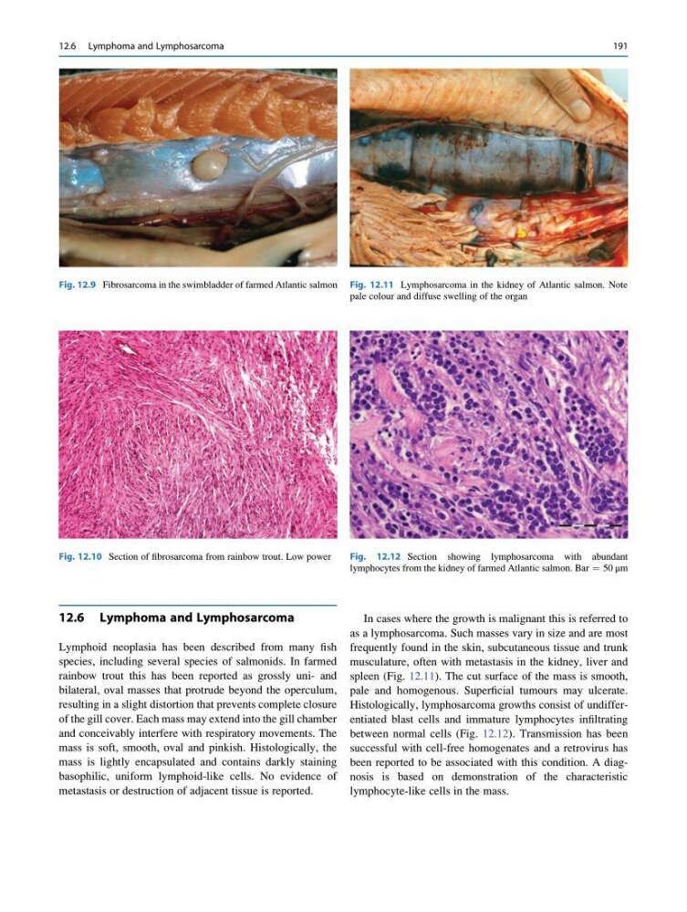
Fig. 12.9 Fibrosarcoma in the swimbladder of farmed Atlantic salmon
Fig. 12.10 Section of fibrosarcoma from rainbow trout. Low power
Fig. 12.11 Lymphosarcoma in the kidney of Atlantic salmon. Note
pale colour and diffuse swelling of the organ
Fig. 12.12 Section showing lymphosarcoma with abundant
lymphocytes from the kidney of farmed Atlantic salmon. Bar ¼ 50 μm
12.6 Lymphoma and Lymphosarcoma
191
Lymphoid neoplasia has been described from many fish
species, including several species of salmonids. In farmed
rainbow trout this has been reported as grossly uni- and
bilateral, oval masses that protrude beyond the operculum,
resulting in a slight distortion that prevents complete closure
of the gill cover. Each mass may extend into the gill chamber
and conceivably interfere with respiratory movements. The
mass is soft, smooth, oval and pinkish. Histologically, the
mass is lightly encapsulated and contains darkly staining
basophilic, uniform lymphoid-like cells. No evidence of
metastasis or destruction of adjacent tissue is reported.
In cases where the growth is malignant this is referred to
as a lymphosarcoma. Such masses vary in size and are most
frequently found in the skin, subcutaneous tissue and trunk
musculature, often with metastasis in the kidney, liver and
spleen (Fig. 12.11). The cut surface of the mass is smooth,
pale and homogenous. Superficial tumours may ulcerate.
Histologically, lymphosarcoma growths consist of undifferentiated blast cells and immature lymphocytes infiltrating
between normal cells (Fig. 12.12). Transmission has been
successful with cell-free homogenates and a retrovirus has
been reported to be associated with this condition. A diagnosis is based on demonstration of the characteristic
lymphocyte-like cells in the mass.
Fig. 12.9 Fibrosarcoma in the swimbladder of farmed Atlantic salmon
Fig. 12.10 Section of fibrosarcoma from rainbow trout. Low power
Fig. 12.11 Lymphosarcoma in the kidney of Atlantic salmon. Note
pale colour and diffuse swelling of the organ
Fig. 12.12 Section showing lymphosarcoma with abundant
lymphocytes from the kidney of farmed Atlantic salmon. Bar ¼ 50 μm
12.6 Lymphoma and Lymphosarcoma
191