

12.4 Leiomyosarcoma
Swim bladder sarcoma (leiomyosarcoma) is infrequently
reported, with a few cases described from maturing seareared Atlantic and wild sockeye salmon. Moribund fish
are generally in poor condition and sluggish, but show no
other external changes. At necropsy, multi-nodular masses
of neoplastic cells occupying external and internal surfaces
of the swim bladder, are randomly scattered. Single, hard
nodular areas to a more extensive encrustment running the
entire length of the organ protrude from the swim bladder
surface into the abdominal cavity (Fig. 12.8). No evidence of
infiltrative growth or distant metastasis has been reported.
Histologically, the neoplasia is well differentiated and
consist of interlacing bundles of spindle cells with round or
elongated nuclei, which arise from the junction of the inner
smooth muscle and the areolar zone of the swim bladder.
These cells have round to oval nuclei with lightly stippled
chromatin, and a single small nucleolus. Some nuclei may
have slight margination of chromatin while other cells show
enlarged irregularly shaped or clefted nuclei and form nests
of anaplastic cells. A whirling arrangement of fibroblasts and
smooth muscle makes up the mass. A novel piscine retrovirus (swim bladder sarcoma virus) has been identified in
association with an occurrence in Atlantic salmon.
The diagnosis of leiomyosarcoma is based on the appearance
of the well differentiated spindle-shaped cells with elongated
cytoplasmic processes, low collagen and high mitotic index.
12.5 Fibrosarcoma
Fibromas and fibrosarcomas are masses of mesenchymal cell
origin that are composed of benign and malignant fibroblasts
and are usually found as nodular, well-defined lesions on or
near the body surface. These masses are soft (myxomas)
with a smooth, pale cut surface. Fibroblasts of variable
differentiation and collagen, often arranged in sworls and
whirls constitute the stroma. Central necrosis in the stroma
may occur with occasional metastases in the kidney and
swim bladder (Fig. 12.9). These neoplasms are readily
distinguished histologically with the demonstration of elongated fibroblasts and dense collagenous fibres in characteristic whirling patterns (Fig. 12.10).
Fig. 12.7 Iridophoroma in the integument of rainbow trout. Low power
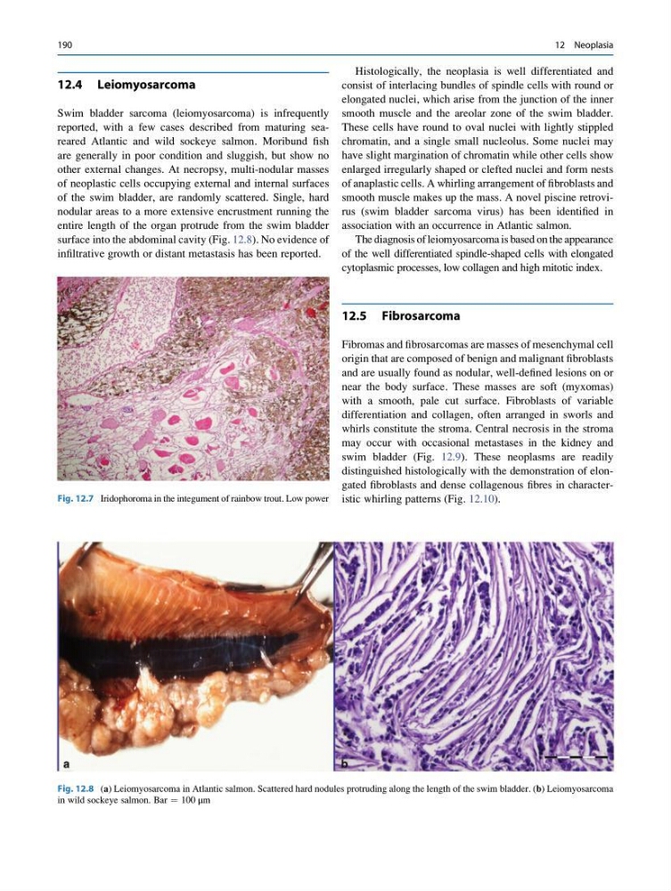
Fig. 12.8 (a) Leiomyosarcoma in Atlantic salmon. Scattered hard nodules protruding along the length of the swim bladder. (b) Leiomyosarcoma
in wild sockeye salmon. Bar ¼ 100 μm
190
12 Neoplasia
Swim bladder sarcoma (leiomyosarcoma) is infrequently
reported, with a few cases described from maturing seareared Atlantic and wild sockeye salmon. Moribund fish
are generally in poor condition and sluggish, but show no
other external changes. At necropsy, multi-nodular masses
of neoplastic cells occupying external and internal surfaces
of the swim bladder, are randomly scattered. Single, hard
nodular areas to a more extensive encrustment running the
entire length of the organ protrude from the swim bladder
surface into the abdominal cavity (Fig. 12.8). No evidence of
infiltrative growth or distant metastasis has been reported.
Histologically, the neoplasia is well differentiated and
consist of interlacing bundles of spindle cells with round or
elongated nuclei, which arise from the junction of the inner
smooth muscle and the areolar zone of the swim bladder.
These cells have round to oval nuclei with lightly stippled
chromatin, and a single small nucleolus. Some nuclei may
have slight margination of chromatin while other cells show
enlarged irregularly shaped or clefted nuclei and form nests
of anaplastic cells. A whirling arrangement of fibroblasts and
smooth muscle makes up the mass. A novel piscine retrovirus (swim bladder sarcoma virus) has been identified in
association with an occurrence in Atlantic salmon.
The diagnosis of leiomyosarcoma is based on the appearance
of the well differentiated spindle-shaped cells with elongated
cytoplasmic processes, low collagen and high mitotic index.
12.5 Fibrosarcoma
Fibromas and fibrosarcomas are masses of mesenchymal cell
origin that are composed of benign and malignant fibroblasts
and are usually found as nodular, well-defined lesions on or
near the body surface. These masses are soft (myxomas)
with a smooth, pale cut surface. Fibroblasts of variable
differentiation and collagen, often arranged in sworls and
whirls constitute the stroma. Central necrosis in the stroma
may occur with occasional metastases in the kidney and
swim bladder (Fig. 12.9). These neoplasms are readily
distinguished histologically with the demonstration of elongated fibroblasts and dense collagenous fibres in characteristic whirling patterns (Fig. 12.10).
Fig. 12.7 Iridophoroma in the integument of rainbow trout. Low power
Fig. 12.8 (a) Leiomyosarcoma in Atlantic salmon. Scattered hard nodules protruding along the length of the swim bladder. (b) Leiomyosarcoma
in wild sockeye salmon. Bar ¼ 100 μm
190
12 Neoplasia