

are natural occurrences, hence the cause or causes are considered to be spontaneous in nature. The topographic localization and histological features are used for diagnosis.
12.3 Pigment Cells
12.3.1 Melanoma
Pigment cell neoplasia is occasionally reported in salmonids,
with melanomas being the most common type from this
group. Both wild and farmed stock may be affected. The
mature growths are generally raised, soft, black-pigmented
areas which are visible on the body surface and underlying
muscle (Fig. 12.5). The neoplasia shows invasion by
melanomacrophages in varying degrees of differentiation,
with fibrous deposition (Fig. 12.6). Metastasis has been
recorded. Gross observations and the cellular detail described from stained histological sections are used to confirm
the diagnosis.
12.3.2 Iridophoroma
Iridophoromas consist of neoplastic iridophores, pigment
fusiform cells which are arranged in bundles with moderate
amount of cytoplasm containing olive to green pigment,
crystalline and birefringent with polarized light. Their nuclei
are round to ovoid with one to two nucleoli and mitotic
figures are not reported. Macroscopically, iridophoromas
comprise a well demarcated, whitish oval mass, raised
above the surface of the skin. The overlying epidermis is
slightly ulcerated with no additional pathological lesions or
metastases reported. Histologically, the mass originates
from the dermal pigment layer and normally encapsulated
(Fig. 12.7). In a few areas the neoplastic cells infiltrate into
the surrounding tissue. The epidermis is normally structured
at the sides of the mass and completely eroded on the surface
of the neoplasia. A severe oedema is reported in the dermis
adjacent to the neoplasia.
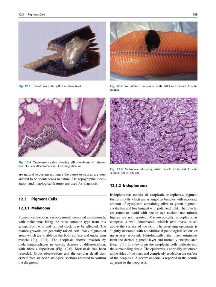
Fig. 12.3 Chondroma in the gill of rainbow trout
Fig. 12.4 Transverse section showing gill chondroma in rainbow
trout. Lillie’s allochrome stain. Low magnification
Fig. 12.5 Well-defined melanoma in the fillet of a farmed Atlantic
salmon
Fig. 12.6 Melanoma infiltrating white muscle of farmed Atlantic
salmon. Bar ¼ 200 μm
12.3 Pigment Cells
189
12.3 Pigment Cells
12.3.1 Melanoma
Pigment cell neoplasia is occasionally reported in salmonids,
with melanomas being the most common type from this
group. Both wild and farmed stock may be affected. The
mature growths are generally raised, soft, black-pigmented
areas which are visible on the body surface and underlying
muscle (Fig. 12.5). The neoplasia shows invasion by
melanomacrophages in varying degrees of differentiation,
with fibrous deposition (Fig. 12.6). Metastasis has been
recorded. Gross observations and the cellular detail described from stained histological sections are used to confirm
the diagnosis.
12.3.2 Iridophoroma
Iridophoromas consist of neoplastic iridophores, pigment
fusiform cells which are arranged in bundles with moderate
amount of cytoplasm containing olive to green pigment,
crystalline and birefringent with polarized light. Their nuclei
are round to ovoid with one to two nucleoli and mitotic
figures are not reported. Macroscopically, iridophoromas
comprise a well demarcated, whitish oval mass, raised
above the surface of the skin. The overlying epidermis is
slightly ulcerated with no additional pathological lesions or
metastases reported. Histologically, the mass originates
from the dermal pigment layer and normally encapsulated
(Fig. 12.7). In a few areas the neoplastic cells infiltrate into
the surrounding tissue. The epidermis is normally structured
at the sides of the mass and completely eroded on the surface
of the neoplasia. A severe oedema is reported in the dermis
adjacent to the neoplasia.
Fig. 12.3 Chondroma in the gill of rainbow trout
Fig. 12.4 Transverse section showing gill chondroma in rainbow
trout. Lillie’s allochrome stain. Low magnification
Fig. 12.5 Well-defined melanoma in the fillet of a farmed Atlantic
salmon
Fig. 12.6 Melanoma infiltrating white muscle of farmed Atlantic
salmon. Bar ¼ 200 μm
12.3 Pigment Cells
189