

chondroma (Fig. 12.3). These are encapsulated with a
lobular growing pattern that originates from the filament
cartilage, possibly as an ingrowth of the surface covering.
Normal squamous, or sometimes hyperplastic epithelium
covers the surface of the mass with some occasional
invaginations into the underlying stroma with increased
mucous cell activity. Within the dermis, there is hypertrophy and some hyperplasia. Many of these growths contain
a number of cystic spaces beneath the epidermis and are
surrounded by a loose fibrous matrix of adipose tissue
containing strands of immature cartilage (Fig. 12.4).
Melanin granules are evenly spread throughout the dermis
with no evidence of inflammation, infiltrative growth or
distant metastasis. Chondrocytes are infrequent with no
mitotic figures.
The absence of infectious agents coupled with the rarity
of chondromas in populations indicates that these growths
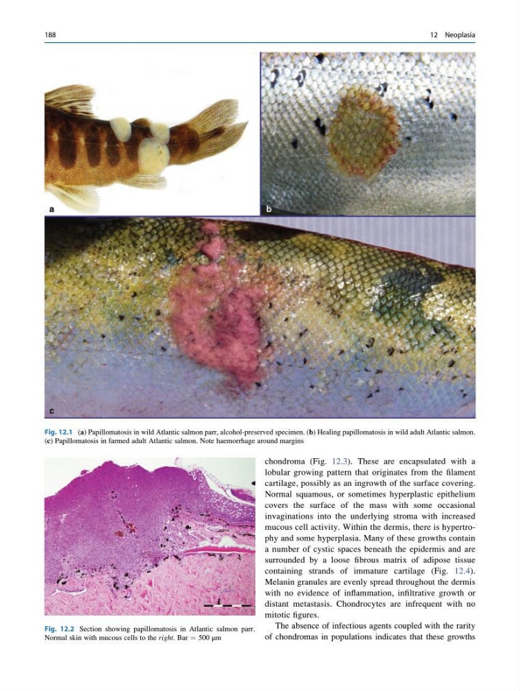
Fig. 12.1 (a) Papillomatosis in wild Atlantic salmon parr, alcohol-preserved specimen. (b) Healing papillomatosis in wild adult Atlantic salmon.
(c) Papillomatosis in farmed adult Atlantic salmon. Note haemorrhage around margins
Fig. 12.2 Section showing papillomatosis in Atlantic salmon parr.
Normal skin with mucous cells to the right. Bar ¼ 500 μm
188
12 Neoplasia
lobular growing pattern that originates from the filament
cartilage, possibly as an ingrowth of the surface covering.
Normal squamous, or sometimes hyperplastic epithelium
covers the surface of the mass with some occasional
invaginations into the underlying stroma with increased
mucous cell activity. Within the dermis, there is hypertrophy and some hyperplasia. Many of these growths contain
a number of cystic spaces beneath the epidermis and are
surrounded by a loose fibrous matrix of adipose tissue
containing strands of immature cartilage (Fig. 12.4).
Melanin granules are evenly spread throughout the dermis
with no evidence of inflammation, infiltrative growth or
distant metastasis. Chondrocytes are infrequent with no
mitotic figures.
The absence of infectious agents coupled with the rarity
of chondromas in populations indicates that these growths
Fig. 12.1 (a) Papillomatosis in wild Atlantic salmon parr, alcohol-preserved specimen. (b) Healing papillomatosis in wild adult Atlantic salmon.
(c) Papillomatosis in farmed adult Atlantic salmon. Note haemorrhage around margins
Fig. 12.2 Section showing papillomatosis in Atlantic salmon parr.
Normal skin with mucous cells to the right. Bar ¼ 500 μm
188
12 Neoplasia