

12.7 Hepatoma
Hepatomas and hepatic carcinomas were recorded in farmed
rainbow trout in the 1960s as a result of Aspergillus flavus
growing on oil seeds during warm and humid storage of the
fish feed, but currently these are rare. Aflatoxins produced
by Aspergillus are highly carcinogenic to rainbow trout and
the affected fish show an enlarged abdomen, splenomegaly
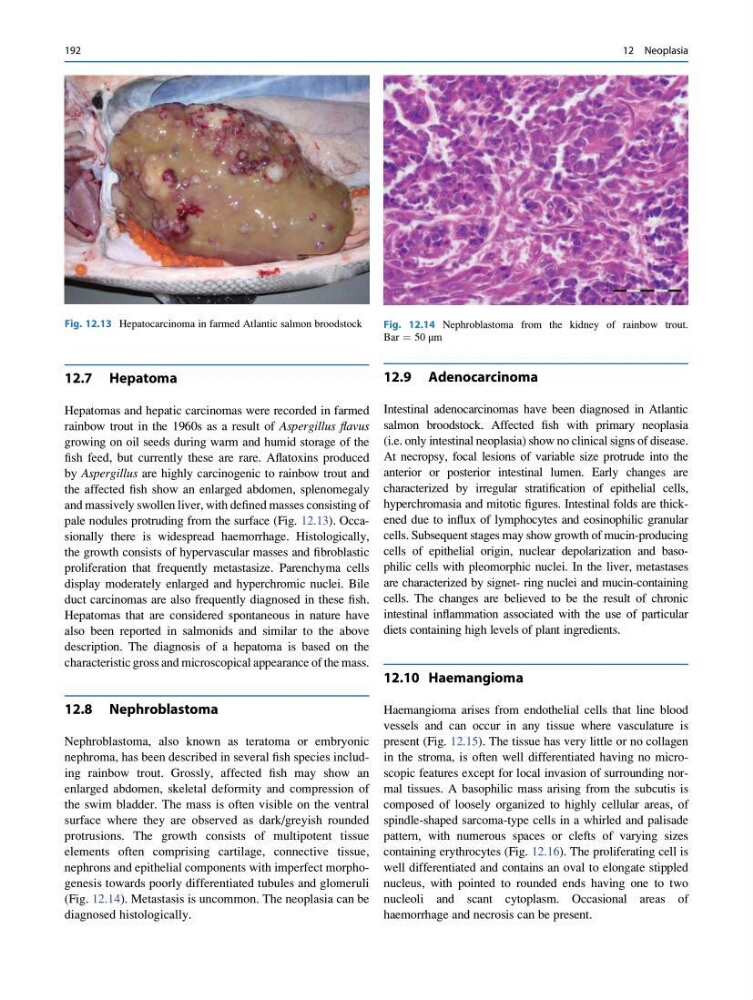
and massively swollen liver, with defined masses consisting of
pale nodules protruding from the surface (Fig. 12.13). Occasionally there is widespread haemorrhage. Histologically,
the growth consists of hypervascular masses and fibroblastic
proliferation that frequently metastasize. Parenchyma cells
display moderately enlarged and hyperchromic nuclei. Bile
duct carcinomas are also frequently diagnosed in these fish.
Hepatomas that are considered spontaneous in nature have
also been reported in salmonids and similar to the above
description. The diagnosis of a hepatoma is based on the
characteristic gross and microscopical appearance of the mass.
12.8 Nephroblastoma
Nephroblastoma, also known as teratoma or embryonic
nephroma, has been described in several fish species including rainbow trout. Grossly, affected fish may show an
enlarged abdomen, skeletal deformity and compression of
the swim bladder. The mass is often visible on the ventral
surface where they are observed as dark/greyish rounded
protrusions. The growth consists of multipotent tissue
elements often comprising cartilage, connective tissue,
nephrons and epithelial components with imperfect morphogenesis towards poorly differentiated tubules and glomeruli
(Fig. 12.14). Metastasis is uncommon. The neoplasia can be
diagnosed histologically.
12.9 Adenocarcinoma
Intestinal adenocarcinomas have been diagnosed in Atlantic
salmon broodstock. Affected fish with primary neoplasia
(i.e. only intestinal neoplasia) show no clinical signs of disease.
At necropsy, focal lesions of variable size protrude into the
anterior or posterior intestinal lumen. Early changes are
characterized by irregular stratification of epithelial cells,
hyperchromasia and mitotic figures. Intestinal folds are thickened due to influx of lymphocytes and eosinophilic granular
cells. Subsequent stages may show growth of mucin-producing
cells of epithelial origin, nuclear depolarization and basophilic cells with pleomorphic nuclei. In the liver, metastases
are characterized by signet- ring nuclei and mucin-containing
cells. The changes are believed to be the result of chronic
intestinal inflammation associated with the use of particular
diets containing high levels of plant ingredients.
12.10 Haemangioma
Haemangioma arises from endothelial cells that line blood
vessels and can occur in any tissue where vasculature is
present (Fig. 12.15). The tissue has very little or no collagen
in the stroma, is often well differentiated having no microscopic features except for local invasion of surrounding normal tissues. A basophilic mass arising from the subcutis is
composed of loosely organized to highly cellular areas, of
spindle-shaped sarcoma-type cells in a whirled and palisade
pattern, with numerous spaces or clefts of varying sizes
containing erythrocytes (Fig. 12.16). The proliferating cell is
well differentiated and contains an oval to elongate stippled
nucleus, with pointed to rounded ends having one to two
nucleoli and scant cytoplasm. Occasional areas of
haemorrhage and necrosis can be present.
Fig. 12.13 Hepatocarcinoma in farmed Atlantic salmon broodstock
Fig. 12.14 Nephroblastoma from the kidney of rainbow trout.
Bar ¼ 50 μm
192
12 Neoplasia
Hepatomas and hepatic carcinomas were recorded in farmed
rainbow trout in the 1960s as a result of Aspergillus flavus
growing on oil seeds during warm and humid storage of the
fish feed, but currently these are rare. Aflatoxins produced
by Aspergillus are highly carcinogenic to rainbow trout and
the affected fish show an enlarged abdomen, splenomegaly
and massively swollen liver, with defined masses consisting of
pale nodules protruding from the surface (Fig. 12.13). Occasionally there is widespread haemorrhage. Histologically,
the growth consists of hypervascular masses and fibroblastic
proliferation that frequently metastasize. Parenchyma cells
display moderately enlarged and hyperchromic nuclei. Bile
duct carcinomas are also frequently diagnosed in these fish.
Hepatomas that are considered spontaneous in nature have
also been reported in salmonids and similar to the above
description. The diagnosis of a hepatoma is based on the
characteristic gross and microscopical appearance of the mass.
12.8 Nephroblastoma
Nephroblastoma, also known as teratoma or embryonic
nephroma, has been described in several fish species including rainbow trout. Grossly, affected fish may show an
enlarged abdomen, skeletal deformity and compression of
the swim bladder. The mass is often visible on the ventral
surface where they are observed as dark/greyish rounded
protrusions. The growth consists of multipotent tissue
elements often comprising cartilage, connective tissue,
nephrons and epithelial components with imperfect morphogenesis towards poorly differentiated tubules and glomeruli
(Fig. 12.14). Metastasis is uncommon. The neoplasia can be
diagnosed histologically.
12.9 Adenocarcinoma
Intestinal adenocarcinomas have been diagnosed in Atlantic
salmon broodstock. Affected fish with primary neoplasia
(i.e. only intestinal neoplasia) show no clinical signs of disease.
At necropsy, focal lesions of variable size protrude into the
anterior or posterior intestinal lumen. Early changes are
characterized by irregular stratification of epithelial cells,
hyperchromasia and mitotic figures. Intestinal folds are thickened due to influx of lymphocytes and eosinophilic granular
cells. Subsequent stages may show growth of mucin-producing
cells of epithelial origin, nuclear depolarization and basophilic cells with pleomorphic nuclei. In the liver, metastases
are characterized by signet- ring nuclei and mucin-containing
cells. The changes are believed to be the result of chronic
intestinal inflammation associated with the use of particular
diets containing high levels of plant ingredients.
12.10 Haemangioma
Haemangioma arises from endothelial cells that line blood
vessels and can occur in any tissue where vasculature is
present (Fig. 12.15). The tissue has very little or no collagen
in the stroma, is often well differentiated having no microscopic features except for local invasion of surrounding normal tissues. A basophilic mass arising from the subcutis is
composed of loosely organized to highly cellular areas, of
spindle-shaped sarcoma-type cells in a whirled and palisade
pattern, with numerous spaces or clefts of varying sizes
containing erythrocytes (Fig. 12.16). The proliferating cell is
well differentiated and contains an oval to elongate stippled
nucleus, with pointed to rounded ends having one to two
nucleoli and scant cytoplasm. Occasional areas of
haemorrhage and necrosis can be present.
Fig. 12.13 Hepatocarcinoma in farmed Atlantic salmon broodstock
Fig. 12.14 Nephroblastoma from the kidney of rainbow trout.
Bar ¼ 50 μm
192
12 Neoplasia