

tissues in affected fish are generally within normal range. The
cause of these conditions is unknown, but the apparent low
prevalence in populations indicates the cysts might be of
congenital origin. The diagnosis of polycystic condition and
similar cystic conditions involving other tissues, is based on
gross observations with additional information from histological assessment of stained sections.
11.4 White Eye Syndrome
White eye syndrome (or generalized soft tissue calcification)
has occurred sporadically in hatcheries and smolt farms in
Norway. Affected fish appear emaciated with exophthalmia
and characteristic crescent-shaped white areas in front and
behind the eye (Fig. 11.11). At necropsy, the body muscle is
speckled with diffuse pale or white patches. Nodular white
lesions may also be found in other organs including heart,
kidney, liver and the gill arches (Fig. 11.12).
Histology reveals calcium deposits, extensive muscle
degeneration and necrosis with calcification of body muscle,
compact and spongy myocardium, cardiac valves and coronary artery. Heavy calcium deposits in the epicardium may
also be found. Mineralized foci are also found in other
organs such as liver, kidney, stomach wall and in the
retrobulbar tissue (corpus choroidale). The aetiology is
unknown, but is probably related to an alteration of the
metabolism of the calcium-regulating mechanisms. Diagnosis is based upon characteristic gross and histological
lesions.
Fig. 11.9 Polycystic liver with cysts of variable size in farmed Atlantic salmon
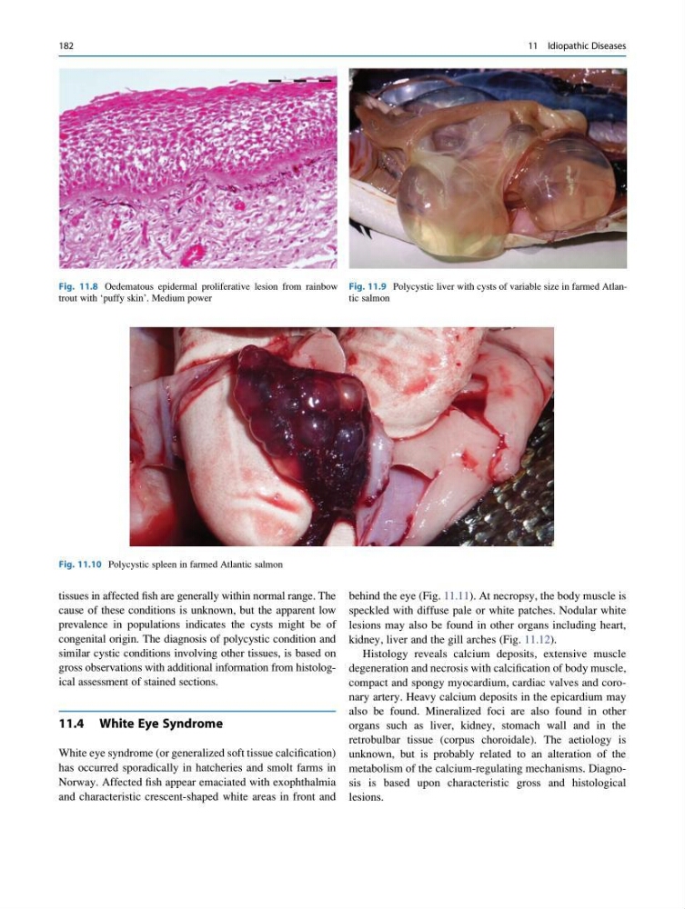
Fig. 11.10 Polycystic spleen in farmed Atlantic salmon
Fig. 11.8 Oedematous epidermal proliferative lesion from rainbow
trout with ‘puffy skin’. Medium power
182
11 Idiopathic Diseases
cause of these conditions is unknown, but the apparent low
prevalence in populations indicates the cysts might be of
congenital origin. The diagnosis of polycystic condition and
similar cystic conditions involving other tissues, is based on
gross observations with additional information from histological assessment of stained sections.
11.4 White Eye Syndrome
White eye syndrome (or generalized soft tissue calcification)
has occurred sporadically in hatcheries and smolt farms in
Norway. Affected fish appear emaciated with exophthalmia
and characteristic crescent-shaped white areas in front and
behind the eye (Fig. 11.11). At necropsy, the body muscle is
speckled with diffuse pale or white patches. Nodular white
lesions may also be found in other organs including heart,
kidney, liver and the gill arches (Fig. 11.12).
Histology reveals calcium deposits, extensive muscle
degeneration and necrosis with calcification of body muscle,
compact and spongy myocardium, cardiac valves and coronary artery. Heavy calcium deposits in the epicardium may
also be found. Mineralized foci are also found in other
organs such as liver, kidney, stomach wall and in the
retrobulbar tissue (corpus choroidale). The aetiology is
unknown, but is probably related to an alteration of the
metabolism of the calcium-regulating mechanisms. Diagnosis is based upon characteristic gross and histological
lesions.
Fig. 11.9 Polycystic liver with cysts of variable size in farmed Atlantic salmon
Fig. 11.10 Polycystic spleen in farmed Atlantic salmon
Fig. 11.8 Oedematous epidermal proliferative lesion from rainbow
trout with ‘puffy skin’. Medium power
182
11 Idiopathic Diseases