

macroscopic characteristics of early skin lesions is considered
pathognomonic for UDN, however, the presence of hyphae
reduces the likelihood of a definitive diagnosis. In the absence
of a clear aetiology and an agreed case definition for this
condition, it is fair to highlight that other skin conditions
may also present some or all of the histological features
described.
11.2 Puffy Skin
‘Puffy skin’ of farmed rainbow trout was first described in
1997 in Scotland and data suggests around 32 sites had
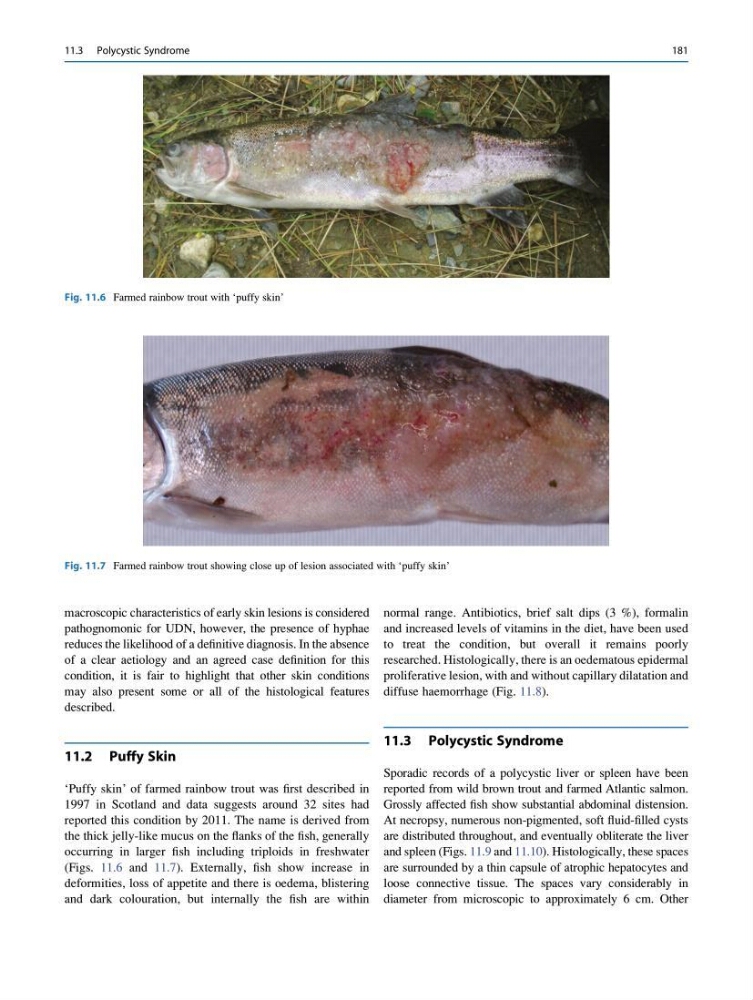
reported this condition by 2011. The name is derived from
the thick jelly-like mucus on the flanks of the fish, generally
occurring in larger fish including triploids in freshwater
(Figs. 11.6 and 11.7). Externally, fish show increase in
deformities, loss of appetite and there is oedema, blistering
and dark colouration, but internally the fish are within
normal range. Antibiotics, brief salt dips (3 %), formalin
and increased levels of vitamins in the diet, have been used
to treat the condition, but overall it remains poorly
researched. Histologically, there is an oedematous epidermal
proliferative lesion, with and without capillary dilatation and
diffuse haemorrhage (Fig. 11.8).
11.3 Polycystic Syndrome
Sporadic records of a polycystic liver or spleen have been
reported from wild brown trout and farmed Atlantic salmon.
Grossly affected fish show substantial abdominal distension.
At necropsy, numerous non-pigmented, soft fluid-filled cysts
are distributed throughout, and eventually obliterate the liver
and spleen (Figs. 11.9 and 11.10). Histologically, these spaces
are surrounded by a thin capsule of atrophic hepatocytes and
loose connective tissue. The spaces vary considerably in
diameter from microscopic to approximately 6 cm. Other
Fig. 11.6 Farmed rainbow trout with ‘puffy skin’
Fig. 11.7 Farmed rainbow trout showing close up of lesion associated with ‘puffy skin’
11.3 Polycystic Syndrome
181
pathognomonic for UDN, however, the presence of hyphae
reduces the likelihood of a definitive diagnosis. In the absence
of a clear aetiology and an agreed case definition for this
condition, it is fair to highlight that other skin conditions
may also present some or all of the histological features
described.
11.2 Puffy Skin
‘Puffy skin’ of farmed rainbow trout was first described in
1997 in Scotland and data suggests around 32 sites had
reported this condition by 2011. The name is derived from
the thick jelly-like mucus on the flanks of the fish, generally
occurring in larger fish including triploids in freshwater
(Figs. 11.6 and 11.7). Externally, fish show increase in
deformities, loss of appetite and there is oedema, blistering
and dark colouration, but internally the fish are within
normal range. Antibiotics, brief salt dips (3 %), formalin
and increased levels of vitamins in the diet, have been used
to treat the condition, but overall it remains poorly
researched. Histologically, there is an oedematous epidermal
proliferative lesion, with and without capillary dilatation and
diffuse haemorrhage (Fig. 11.8).
11.3 Polycystic Syndrome
Sporadic records of a polycystic liver or spleen have been
reported from wild brown trout and farmed Atlantic salmon.
Grossly affected fish show substantial abdominal distension.
At necropsy, numerous non-pigmented, soft fluid-filled cysts
are distributed throughout, and eventually obliterate the liver
and spleen (Figs. 11.9 and 11.10). Histologically, these spaces
are surrounded by a thin capsule of atrophic hepatocytes and
loose connective tissue. The spaces vary considerably in
diameter from microscopic to approximately 6 cm. Other
Fig. 11.6 Farmed rainbow trout with ‘puffy skin’
Fig. 11.7 Farmed rainbow trout showing close up of lesion associated with ‘puffy skin’
11.3 Polycystic Syndrome
181