

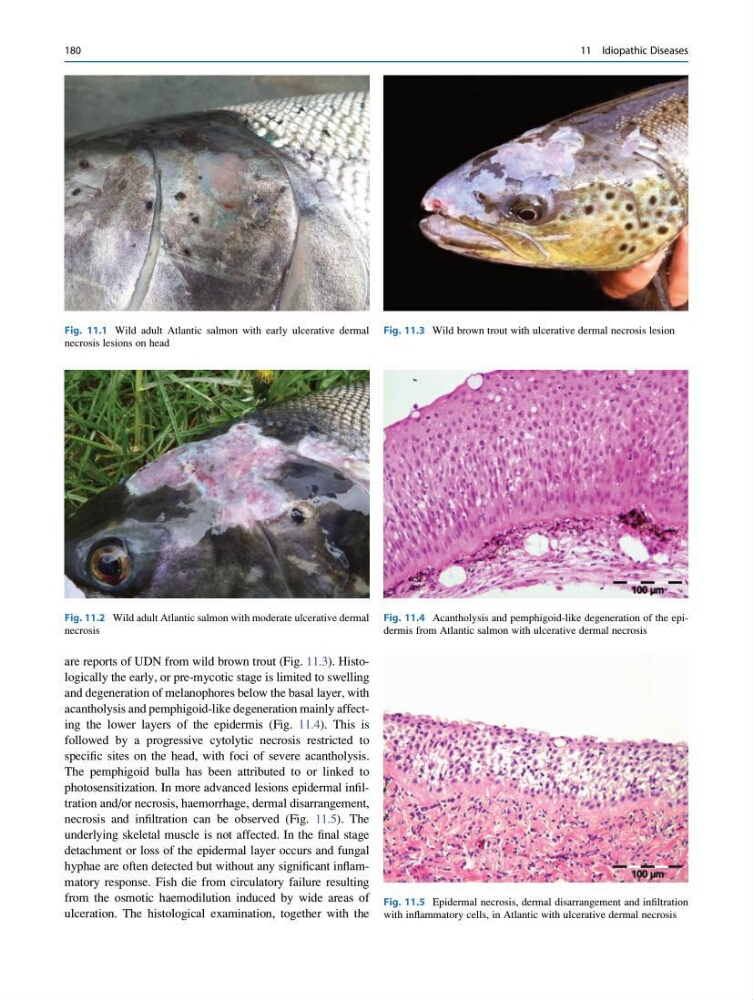
are reports of UDN from wild brown trout (Fig. 11.3). Histologically the early, or pre-mycotic stage is limited to swelling
and degeneration of melanophores below the basal layer, with
acantholysis and pemphigoid-like degeneration mainly affecting the lower layers of the epidermis (Fig. 11.4). This is
followed by a progressive cytolytic necrosis restricted to
specific sites on the head, with foci of severe acantholysis.
The pemphigoid bulla has been attributed to or linked to
photosensitization. In more advanced lesions epidermal infiltration and/or necrosis, haemorrhage, dermal disarrangement,
necrosis and infiltration can be observed (Fig. 11.5). The
underlying skeletal muscle is not affected. In the final stage
detachment or loss of the epidermal layer occurs and fungal
hyphae are often detected but without any significant inflammatory response. Fish die from circulatory failure resulting
from the osmotic haemodilution induced by wide areas of
ulceration. The histological examination, together with the
Fig. 11.1 Wild adult Atlantic salmon with early ulcerative dermal
necrosis lesions on head
Fig. 11.2 Wild adult Atlantic salmon with moderate ulcerative dermal
necrosis
Fig. 11.3 Wild brown trout with ulcerative dermal necrosis lesion
Fig. 11.4 Acantholysis and pemphigoid-like degeneration of the epidermis from Atlantic salmon with ulcerative dermal necrosis
Fig. 11.5 Epidermal necrosis, dermal disarrangement and infiltration
with inflammatory cells, in Atlantic with ulcerative dermal necrosis
180
11 Idiopathic Diseases
and degeneration of melanophores below the basal layer, with
acantholysis and pemphigoid-like degeneration mainly affecting the lower layers of the epidermis (Fig. 11.4). This is
followed by a progressive cytolytic necrosis restricted to
specific sites on the head, with foci of severe acantholysis.
The pemphigoid bulla has been attributed to or linked to
photosensitization. In more advanced lesions epidermal infiltration and/or necrosis, haemorrhage, dermal disarrangement,
necrosis and infiltration can be observed (Fig. 11.5). The
underlying skeletal muscle is not affected. In the final stage
detachment or loss of the epidermal layer occurs and fungal
hyphae are often detected but without any significant inflammatory response. Fish die from circulatory failure resulting
from the osmotic haemodilution induced by wide areas of
ulceration. The histological examination, together with the
Fig. 11.1 Wild adult Atlantic salmon with early ulcerative dermal
necrosis lesions on head
Fig. 11.2 Wild adult Atlantic salmon with moderate ulcerative dermal
necrosis
Fig. 11.3 Wild brown trout with ulcerative dermal necrosis lesion
Fig. 11.4 Acantholysis and pemphigoid-like degeneration of the epidermis from Atlantic salmon with ulcerative dermal necrosis
Fig. 11.5 Epidermal necrosis, dermal disarrangement and infiltration
with inflammatory cells, in Atlantic with ulcerative dermal necrosis
180
11 Idiopathic Diseases