

endocardium and the cardiac myofibres and can be identified
using Elastin-van Gieson staining. Lesions may be found in
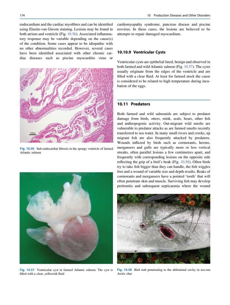
both atrium and ventricle (Fig. 10.56). Associated inflammatory response may be variable depending on the cause(s)
of the condition. Some cases appear to be idiopathic with
no other abnormalities recorded. However, several cases
have been identified associated with other chronic cardiac diseases such as piscine myocarditis virus or
cardiomyopathy syndrome, pancreas disease and piscine
reovirus. In these cases, the lesions are believed to be
attempts to repair damaged myocardium.
10.10.9 Ventricular Cysts
Ventricular cysts are epithelial lined, benign and observed in
both farmed and wild Atlantic salmon (Fig. 10.57). The cysts
usually originate from the edges of the ventricle and are
filled with a clear fluid. At least for farmed stock the cause
is considered to be related to high temperature during incubation of the eggs.
10.11 Predators
Both farmed and wild salmonids are subject to predator
damage from birds, otters, mink, seals, bears, other fish
and anthropogenic activity. Out-migrant wild smolts are
vulnerable to predator attacks as are farmed smolts recently
transferred to sea water. In many small rivers and creeks, up
migrant fish are also frequently attacked by predators.
Wounds inflicted by birds such as cormorants, herons,
mergansers and gulls are typically more or less vertical
streaks, often parallel lesions a few centimetres apart, and
frequently with corresponding lesions on the opposite side
reflecting the grip of a bird’s beak (Fig. 10.58). Often birds
try to take fish bigger than they can handle, the fish wiggles
free and a wound of variable size and depth results. Beaks of
cormorants and mergansers have a pointed ‘tooth’ that will
often penetrate skin and muscle. Surviving fish may develop
peritonitis and subsequent septicaemia where the wound
Fig. 10.56 Sub-endocardial fibrosis in the spongy ventricle of farmed
Atlantic salmon
Fig. 10.57 Ventricular cyst in farmed Atlantic salmon. The cyst is
filled with a clear, yellowish fluid
Fig. 10.58 Bird stab penetrating to the abdominal cavity in sea-run
Arctic char
174
10 Production Diseases and Other Disorders
using Elastin-van Gieson staining. Lesions may be found in
both atrium and ventricle (Fig. 10.56). Associated inflammatory response may be variable depending on the cause(s)
of the condition. Some cases appear to be idiopathic with
no other abnormalities recorded. However, several cases
have been identified associated with other chronic cardiac diseases such as piscine myocarditis virus or
cardiomyopathy syndrome, pancreas disease and piscine
reovirus. In these cases, the lesions are believed to be
attempts to repair damaged myocardium.
10.10.9 Ventricular Cysts
Ventricular cysts are epithelial lined, benign and observed in
both farmed and wild Atlantic salmon (Fig. 10.57). The cysts
usually originate from the edges of the ventricle and are
filled with a clear fluid. At least for farmed stock the cause
is considered to be related to high temperature during incubation of the eggs.
10.11 Predators
Both farmed and wild salmonids are subject to predator
damage from birds, otters, mink, seals, bears, other fish
and anthropogenic activity. Out-migrant wild smolts are
vulnerable to predator attacks as are farmed smolts recently
transferred to sea water. In many small rivers and creeks, up
migrant fish are also frequently attacked by predators.
Wounds inflicted by birds such as cormorants, herons,
mergansers and gulls are typically more or less vertical
streaks, often parallel lesions a few centimetres apart, and
frequently with corresponding lesions on the opposite side
reflecting the grip of a bird’s beak (Fig. 10.58). Often birds
try to take fish bigger than they can handle, the fish wiggles
free and a wound of variable size and depth results. Beaks of
cormorants and mergansers have a pointed ‘tooth’ that will
often penetrate skin and muscle. Surviving fish may develop
peritonitis and subsequent septicaemia where the wound
Fig. 10.56 Sub-endocardial fibrosis in the spongy ventricle of farmed
Atlantic salmon
Fig. 10.57 Ventricular cyst in farmed Atlantic salmon. The cyst is
filled with a clear, yellowish fluid
Fig. 10.58 Bird stab penetrating to the abdominal cavity in sea-run
Arctic char
174
10 Production Diseases and Other Disorders