

these abnormalities will always be impaired swimming capability, hence more energy is used for locomotion and feeding
and therefore vertebral column abnormalities lesions are often
protracted. Such fish have reduced stress tolerance, are more
susceptible to physiological imbalance and are consequently
always inferior to fish with normal function. Wild fish with
such lesions will typically be poorer in the competition for
food and territory and will also be readily predated.
The aetiology may be complex including congenital (e.g.
inbreeding, genetic factors), idiopathic or acquired (e.g. infectious, nutritional or physical). Under farming conditions, vertebral column abnormalities has been associated alone or in
combination, to different factors such as parasitic infections,
nutritional deficiency and imbalance (e.g. phosphorous and
vitamin C), vaccination, elevated temperature (during egg
incubation and yolk-sac period), adverse environmental
conditions such as hypoxia, exposure to toxicants, fast growth,
or obstruction of the pneumatic duct.
Vertebral column malformations are commonly
diagnosed by gross signs and X-ray, while the exact
aetiology may be a greater challenge to solve. An exemption
might be the parasite aetiology, where examination of
homogenized cartilage and gill arches are needed to exclude
Myxobolus spores (see Fig. 9.10)
10.9 Congenital Malformations
Congenital malformations occur in both wild and farmed
progeny of salmonid fish and different types may occur
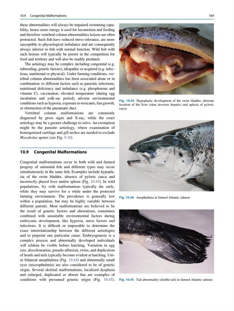
simultaneously in the same fish. Examples include hypoplasia of the swim bladder, absence of pyloric caeca and
incorrectly placed liver and/or spleen (Fig. 10.43). In wild
populations, fry with malformations typically die early,
while they may survive for a while under the protected
farming environment. The prevalence is generally low
within a population, but may be highly variable between
different parents. Most malformations are believed to be
the result of genetic factors and aberrations, sometimes
combined with unsuitable environmental factors during
embryonic development, like hypoxia, stress factors and
infections. It is difficult or impossible to determine the
exact interrelationship between the different aetiologies
and to pinpoint one particular cause. Embryogenesis is a
complex process and abnormally developed individuals
will seldom be visible before hatching. Variation in egg
size, discolouration, pseudo-albinism, twins, and duplication
of heads and tails typically become evident at hatching. Unior bilateral anopthalmia (Fig. 10.44) and abnormally small
eyes (micropthalmia) are also considered to be of genetic
origin. Several skeletal malformations, localized dysplasia
and enlarged, duplicated or absent fins are examples of
conditions with presumed genetic origin (Fig. 10.45).
Fig. 10.43 Hypoplastic development of the swim bladder, aberrant
location of the liver (situs inversus hepatis) and aplasia of pyloric
caeca
Fig. 10.44 Anopthalmia in farmed Atlantic salmon
Fig. 10.45 Tail abnormality (double tail) in farmed Atlantic salmon
10.9 Congenital Malformations
169
and therefore vertebral column abnormalities lesions are often
protracted. Such fish have reduced stress tolerance, are more
susceptible to physiological imbalance and are consequently
always inferior to fish with normal function. Wild fish with
such lesions will typically be poorer in the competition for
food and territory and will also be readily predated.
The aetiology may be complex including congenital (e.g.
inbreeding, genetic factors), idiopathic or acquired (e.g. infectious, nutritional or physical). Under farming conditions, vertebral column abnormalities has been associated alone or in
combination, to different factors such as parasitic infections,
nutritional deficiency and imbalance (e.g. phosphorous and
vitamin C), vaccination, elevated temperature (during egg
incubation and yolk-sac period), adverse environmental
conditions such as hypoxia, exposure to toxicants, fast growth,
or obstruction of the pneumatic duct.
Vertebral column malformations are commonly
diagnosed by gross signs and X-ray, while the exact
aetiology may be a greater challenge to solve. An exemption
might be the parasite aetiology, where examination of
homogenized cartilage and gill arches are needed to exclude
Myxobolus spores (see Fig. 9.10)
10.9 Congenital Malformations
Congenital malformations occur in both wild and farmed
progeny of salmonid fish and different types may occur
simultaneously in the same fish. Examples include hypoplasia of the swim bladder, absence of pyloric caeca and
incorrectly placed liver and/or spleen (Fig. 10.43). In wild
populations, fry with malformations typically die early,
while they may survive for a while under the protected
farming environment. The prevalence is generally low
within a population, but may be highly variable between
different parents. Most malformations are believed to be
the result of genetic factors and aberrations, sometimes
combined with unsuitable environmental factors during
embryonic development, like hypoxia, stress factors and
infections. It is difficult or impossible to determine the
exact interrelationship between the different aetiologies
and to pinpoint one particular cause. Embryogenesis is a
complex process and abnormally developed individuals
will seldom be visible before hatching. Variation in egg
size, discolouration, pseudo-albinism, twins, and duplication
of heads and tails typically become evident at hatching. Unior bilateral anopthalmia (Fig. 10.44) and abnormally small
eyes (micropthalmia) are also considered to be of genetic
origin. Several skeletal malformations, localized dysplasia
and enlarged, duplicated or absent fins are examples of
conditions with presumed genetic origin (Fig. 10.45).
Fig. 10.43 Hypoplastic development of the swim bladder, aberrant
location of the liver (situs inversus hepatis) and aplasia of pyloric
caeca
Fig. 10.44 Anopthalmia in farmed Atlantic salmon
Fig. 10.45 Tail abnormality (double tail) in farmed Atlantic salmon
10.9 Congenital Malformations
169