

occasionally round with a smooth, non-pigmented surface
and measuring up to 1.5 mm in diameter. Mortality
attributed to this defect has ranged from 10 to 15 % in certain
egg batches. Growth of the surviving fry is not impaired and
behaviour appears normal.
Histologically the cerebellum appears normal, but
displaced dorsally. Karyorhectic Malpighian cells are absent
from the epidermis and the meninges appear normal. The
molecular and granular layers of the cerebellum are displaced
upwards towards the frontal plate with no inflammatory reaction or changes in the Purkinje cell layer. There is no apparent
contact of the cerebellum with the water, or evidence of
infection. The eye has been shown to be affected, with the
retina appearing excessively folded with a decrease in the
volume of the vitreous chamber. The aetiology of this condition is currently unknown. Visual observation and histology
are used to identify these nodules.
Jaw (mandible) deformities have been documented in wild
fish but frequencies are greater in hatchery populations.
Deformities can affect both the maxilla and/or the mandible,
resulting in a short or long lower jaw deformity. The latter can
also be displaced laterally. These abnormalities can be induced
during embryonic and post-embryonic periods of life and are
often lethal with over 80 % of the affected larvae dying.
Further causes of deformities have been attributed to a
genetic or environmental origin, the result of adverse environmental changes, phosphorous deficiency, excessive cartilage deposition and physical injury.
10.7.1 Mandibular Ankylosis
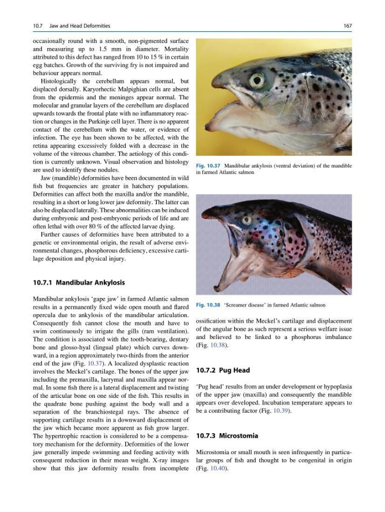
Mandibular ankylosis ‘gape jaw’ in farmed Atlantic salmon
results in a permanently fixed wide open mouth and flared
opercula due to ankylosis of the mandibular articulation.
Consequently fish cannot close the mouth and have to
swim continuously to irrigate the gills (ram ventilation).
The condition is associated with the tooth-bearing, dentary
bone and glosso-hyal (lingual plate) which curves downward, in a region approximately two-thirds from the anterior
end of the jaw (Fig. 10.37). A localized dysplastic reaction
involves the Meckel’s cartilage. The bones of the upper jaw
including the premaxilla, lacrymal and maxilla appear normal. In some fish there is a lateral displacement and twisting
of the articular bone on one side of the fish. This results in
the quadrate bone pushing against the body wall and a
separation of the branchiostegal rays. The absence of
supporting cartilage results in a downward displacement of
the jaw which became more apparent as fish grow larger.
The hypertrophic reaction is considered to be a compensatory mechanism for the deformity. Deformities of the lower
jaw generally impede swimming and feeding activity with
consequent reduction in their mean weight. X-ray images
show that this jaw deformity results from incomplete
ossification within the Meckel’s cartilage and displacement
of the angular bone as such represent a serious welfare issue
and believed to be linked to a phosphorus imbalance
(Fig. 10.38).
10.7.2 Pug Head
‘Pug head’ results from an under development or hypoplasia
of the upper jaw (maxilla) and consequently the mandible
appears over developed. Incubation temperature appears to
be a contributing factor (Fig. 10.39).
10.7.3 Microstomia
Microstomia or small mouth is seen infrequently in particular groups of fish and thought to be congenital in origin
(Fig. 10.40).
Fig. 10.37 Mandibular ankylosis (ventral deviation) of the mandible
in farmed Atlantic salmon
Fig. 10.38 ‘Screamer disease’ in farmed Atlantic salmon
10.7 Jaw and Head Deformities
167
and measuring up to 1.5 mm in diameter. Mortality
attributed to this defect has ranged from 10 to 15 % in certain
egg batches. Growth of the surviving fry is not impaired and
behaviour appears normal.
Histologically the cerebellum appears normal, but
displaced dorsally. Karyorhectic Malpighian cells are absent
from the epidermis and the meninges appear normal. The
molecular and granular layers of the cerebellum are displaced
upwards towards the frontal plate with no inflammatory reaction or changes in the Purkinje cell layer. There is no apparent
contact of the cerebellum with the water, or evidence of
infection. The eye has been shown to be affected, with the
retina appearing excessively folded with a decrease in the
volume of the vitreous chamber. The aetiology of this condition is currently unknown. Visual observation and histology
are used to identify these nodules.
Jaw (mandible) deformities have been documented in wild
fish but frequencies are greater in hatchery populations.
Deformities can affect both the maxilla and/or the mandible,
resulting in a short or long lower jaw deformity. The latter can
also be displaced laterally. These abnormalities can be induced
during embryonic and post-embryonic periods of life and are
often lethal with over 80 % of the affected larvae dying.
Further causes of deformities have been attributed to a
genetic or environmental origin, the result of adverse environmental changes, phosphorous deficiency, excessive cartilage deposition and physical injury.
10.7.1 Mandibular Ankylosis
Mandibular ankylosis ‘gape jaw’ in farmed Atlantic salmon
results in a permanently fixed wide open mouth and flared
opercula due to ankylosis of the mandibular articulation.
Consequently fish cannot close the mouth and have to
swim continuously to irrigate the gills (ram ventilation).
The condition is associated with the tooth-bearing, dentary
bone and glosso-hyal (lingual plate) which curves downward, in a region approximately two-thirds from the anterior
end of the jaw (Fig. 10.37). A localized dysplastic reaction
involves the Meckel’s cartilage. The bones of the upper jaw
including the premaxilla, lacrymal and maxilla appear normal. In some fish there is a lateral displacement and twisting
of the articular bone on one side of the fish. This results in
the quadrate bone pushing against the body wall and a
separation of the branchiostegal rays. The absence of
supporting cartilage results in a downward displacement of
the jaw which became more apparent as fish grow larger.
The hypertrophic reaction is considered to be a compensatory mechanism for the deformity. Deformities of the lower
jaw generally impede swimming and feeding activity with
consequent reduction in their mean weight. X-ray images
show that this jaw deformity results from incomplete
ossification within the Meckel’s cartilage and displacement
of the angular bone as such represent a serious welfare issue
and believed to be linked to a phosphorus imbalance
(Fig. 10.38).
10.7.2 Pug Head
‘Pug head’ results from an under development or hypoplasia
of the upper jaw (maxilla) and consequently the mandible
appears over developed. Incubation temperature appears to
be a contributing factor (Fig. 10.39).
10.7.3 Microstomia
Microstomia or small mouth is seen infrequently in particular groups of fish and thought to be congenital in origin
(Fig. 10.40).
Fig. 10.37 Mandibular ankylosis (ventral deviation) of the mandible
in farmed Atlantic salmon
Fig. 10.38 ‘Screamer disease’ in farmed Atlantic salmon
10.7 Jaw and Head Deformities
167