

unfavourable conditions (e.g. heat, light, moisture) may
compromise the quality and inconsistency may occasionally
occur. In welfare terms there is also increasing evidence that
intensively reared fish significantly alters some aspect of
cardiac anatomy and physiology. In this section the effect of
poor or deficient diets on the health of farmed fish is
discussed.
10.3.1 Inadequate Digestion of Pellet
Dry pellets will normally decompose to a porridge-like
consistency in the stomach before passing to the pyloric
region for further digestion, but at low temperatures, the
feed is not always dissolved in the stomach and may become
lodged as a hard plug in the sphincter area with resultant
accumulation of dry and hard pellets in the stomach
(Fig. 10.10). The mucosa is typically hyperaemic and
irritated, which is also reflected in histopathological lesions.
Another condition that occurs under similar circumstances
is where faeces of farmed salmon turn yellowish and foamy
and float to the surface. The posterior gut is hyperaemic and
the vent opening may be swollen. However, the direct causes
of both conditions are unknown, but are believed to be
associated to the technical and biological qualities of the
pellet at variable ambient temperatures.
10.3.2 Hepatic Lipidosis
Farmed salmonids are susceptible to hepatic lipidosis or
lipoid liver disease (LLD) when fed polyunsaturated fatty
acids combined with insufficient amounts of antioxidant
protection, such as vitamin E. Polyunsaturated fatty acids
are prone to auto-oxidation on exposure to atmospheric
oxygen.
Clinical signs include reduced appetite, impaired equilibrium and increased mortality. Fish become anaemic with
pale gills and ascites, loose scales and develop fin necrosis.
Internally, a yellow-orange discolouration and slight
enlargement of the liver with a friable fatty consistency has
been observed. The haematocrit is usually severely
depressed as a result of a microcytic anaemia with increased
blood cell fragility. Mortalities result from the cumulative
effects of anaemia and hepatocellular and biliary tract dysfunction. Typical histological lesions show accumulation of
excessive lipid in hepatocytes, macrophages and fat-storing
cells (Fig. 10.11). Degenerating hepatocytes with pyknotic
nuclei may occur over large areas. Red cell breakdown
frequently results in haemosiderosis in the spleen. Diagnosis
is based on internal appearance and evidence of a generalised fatty infiltration of the liver in stained tissue sections.
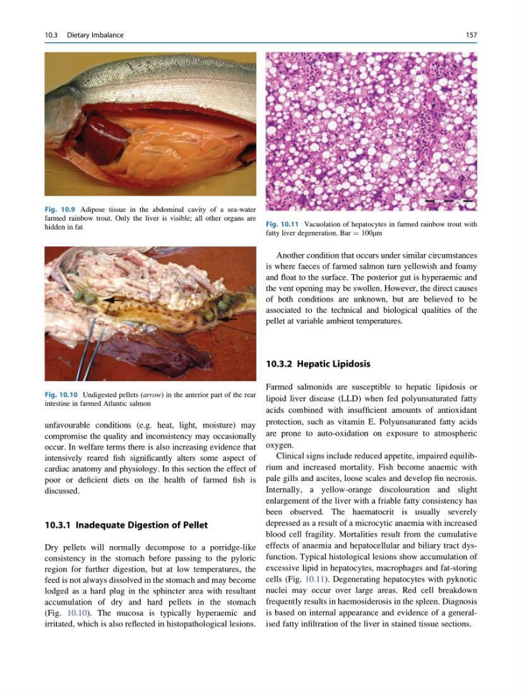
Fig. 10.9 Adipose tissue in the abdominal cavity of a sea-water
farmed rainbow trout. Only the liver is visible; all other organs are
hidden in fat
Fig. 10.10 Undigested pellets (arrow) in the anterior part of the rear
intestine in farmed Atlantic salmon
Fig. 10.11 Vacuolation of hepatocytes in farmed rainbow trout with
fatty liver degeneration. Bar ¼ 100μm
10.3 Dietary Imbalance
157
compromise the quality and inconsistency may occasionally
occur. In welfare terms there is also increasing evidence that
intensively reared fish significantly alters some aspect of
cardiac anatomy and physiology. In this section the effect of
poor or deficient diets on the health of farmed fish is
discussed.
10.3.1 Inadequate Digestion of Pellet
Dry pellets will normally decompose to a porridge-like
consistency in the stomach before passing to the pyloric
region for further digestion, but at low temperatures, the
feed is not always dissolved in the stomach and may become
lodged as a hard plug in the sphincter area with resultant
accumulation of dry and hard pellets in the stomach
(Fig. 10.10). The mucosa is typically hyperaemic and
irritated, which is also reflected in histopathological lesions.
Another condition that occurs under similar circumstances
is where faeces of farmed salmon turn yellowish and foamy
and float to the surface. The posterior gut is hyperaemic and
the vent opening may be swollen. However, the direct causes
of both conditions are unknown, but are believed to be
associated to the technical and biological qualities of the
pellet at variable ambient temperatures.
10.3.2 Hepatic Lipidosis
Farmed salmonids are susceptible to hepatic lipidosis or
lipoid liver disease (LLD) when fed polyunsaturated fatty
acids combined with insufficient amounts of antioxidant
protection, such as vitamin E. Polyunsaturated fatty acids
are prone to auto-oxidation on exposure to atmospheric
oxygen.
Clinical signs include reduced appetite, impaired equilibrium and increased mortality. Fish become anaemic with
pale gills and ascites, loose scales and develop fin necrosis.
Internally, a yellow-orange discolouration and slight
enlargement of the liver with a friable fatty consistency has
been observed. The haematocrit is usually severely
depressed as a result of a microcytic anaemia with increased
blood cell fragility. Mortalities result from the cumulative
effects of anaemia and hepatocellular and biliary tract dysfunction. Typical histological lesions show accumulation of
excessive lipid in hepatocytes, macrophages and fat-storing
cells (Fig. 10.11). Degenerating hepatocytes with pyknotic
nuclei may occur over large areas. Red cell breakdown
frequently results in haemosiderosis in the spleen. Diagnosis
is based on internal appearance and evidence of a generalised fatty infiltration of the liver in stained tissue sections.
Fig. 10.9 Adipose tissue in the abdominal cavity of a sea-water
farmed rainbow trout. Only the liver is visible; all other organs are
hidden in fat
Fig. 10.10 Undigested pellets (arrow) in the anterior part of the rear
intestine in farmed Atlantic salmon
Fig. 10.11 Vacuolation of hepatocytes in farmed rainbow trout with
fatty liver degeneration. Bar ¼ 100μm
10.3 Dietary Imbalance
157