

spawning Atlantic salmon and sea-trout, has been recorded
in Norway. Infected trout may show fibrogranulomatosis of
the epicardium. Diagnosis can be made by clinical signs and
light microscopy.
9.4.6 Cotylurus spp.
There are several species of Cotylurus that infect fish. The
eggs are excreted by fish-eating birds including cormorants,
herons and gulls, and hatch after around 15–16 days. The
circulatory system and loose connective tissue serve as
migratory routes of the metacercaria into the pericardium,
where it encysts in the pericardial cavity of rainbow trout
and Arctic grayling (Fig. 9.40). Overall, there is typically
little tissue response around the encysted metacercariae
although for some species, there may be severe
haemorrhagic enteritis. In heavily infected fish Cotylurus
cause a loss of condition factor and an increased susceptibility to other diseases.
9.4.7 Ichthyocotylurus erraticus
Ichthyocotylurus has a similar life cycle to Apatemon and
Cotylurus. Aggregates of encysted metacercariae of
I. erraticus occur around the bulbus arteriosus and to some
extent also in the epicardium and myocardium forming a
reactive fibro-connective capsule around the parasite in fish
such as grayling and whitefish (Fig. 9.41).
Fish may show skin haemorrhage, especially on the ventral side with raised scales. The pericardium can be covered
with several layers of metacercarial cysts and the mass of
parasites extending deep into the compact layer of the heart;
a PAS-positive reaction in tissue sections helps identify the
cysts walls. A heavy inflammatory reaction occurs around
the parasites. Mononuclear cells (presumably lymphocytes)
and EGCs are the most prevalent cell types in the reaction.
Chronic granuloma formation occurs around the parasites.
The diagnosis is based upon gross, histological findings and
PCR identification of the parasite.
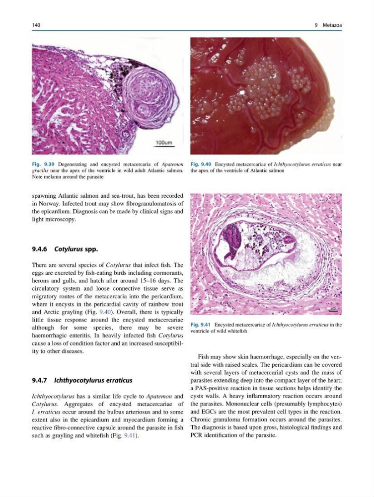
Fig. 9.39 Degenerating and encysted metacercaria of Apatemon
gracilis near the apex of the ventricle in wild adult Atlantic salmon.
Note melanin around the parasite
Fig. 9.40 Encysted metacercariae of Ichthyocotylurus erraticus near
the apex of the ventricle of Atlantic salmon
Fig. 9.41 Encysted metacercariae of Ichthyocotylurus erraticus in the
ventricle of wild whitefish
140
9 Metazoa
in Norway. Infected trout may show fibrogranulomatosis of
the epicardium. Diagnosis can be made by clinical signs and
light microscopy.
9.4.6 Cotylurus spp.
There are several species of Cotylurus that infect fish. The
eggs are excreted by fish-eating birds including cormorants,
herons and gulls, and hatch after around 15–16 days. The
circulatory system and loose connective tissue serve as
migratory routes of the metacercaria into the pericardium,
where it encysts in the pericardial cavity of rainbow trout
and Arctic grayling (Fig. 9.40). Overall, there is typically
little tissue response around the encysted metacercariae
although for some species, there may be severe
haemorrhagic enteritis. In heavily infected fish Cotylurus
cause a loss of condition factor and an increased susceptibility to other diseases.
9.4.7 Ichthyocotylurus erraticus
Ichthyocotylurus has a similar life cycle to Apatemon and
Cotylurus. Aggregates of encysted metacercariae of
I. erraticus occur around the bulbus arteriosus and to some
extent also in the epicardium and myocardium forming a
reactive fibro-connective capsule around the parasite in fish
such as grayling and whitefish (Fig. 9.41).
Fish may show skin haemorrhage, especially on the ventral side with raised scales. The pericardium can be covered
with several layers of metacercarial cysts and the mass of
parasites extending deep into the compact layer of the heart;
a PAS-positive reaction in tissue sections helps identify the
cysts walls. A heavy inflammatory reaction occurs around
the parasites. Mononuclear cells (presumably lymphocytes)
and EGCs are the most prevalent cell types in the reaction.
Chronic granuloma formation occurs around the parasites.
The diagnosis is based upon gross, histological findings and
PCR identification of the parasite.
Fig. 9.39 Degenerating and encysted metacercaria of Apatemon
gracilis near the apex of the ventricle in wild adult Atlantic salmon.
Note melanin around the parasite
Fig. 9.40 Encysted metacercariae of Ichthyocotylurus erraticus near
the apex of the ventricle of Atlantic salmon
Fig. 9.41 Encysted metacercariae of Ichthyocotylurus erraticus in the
ventricle of wild whitefish
140
9 Metazoa