

plerocercoids are found free or encysted on the viscera or in
the musculature, where they can burrow deeply. Commonly,
the plerocercoids trigger a granulomatous encystment on the
viscera, with a dense fibrous coat incorporating fibroblasts,
macrophages and collagenous tissue, and adhesions between
viscera and the abdominal wall can be observed. In the
muscle block plerocercoids may also cause granulomas,
haemorrhage and necrosis with cavitation, negatively affecting the swimming ability of the fish. Plerocercoids are long
lived in the fish intermediate host and will therefore accumulate in older fish that typically develop the most severe
lesions. Piscivorous birds or mammals are final hosts where
reproduction occurs in the gut.
When fish are the final host, adult worms are typically
found attached by their scolex to the intestinal mucosa of
the pyloric caeca and hindgut. Cestoda lack a digestive system
and semi-digested nutrients from the host are absorbed
through the whole body surface. The strobila is composed of
individual proglottids where each segment contains a single
set of reproductive organs. The proglottids with fertilized
eggs are shed from the posterior end of the parasite as
they mature. Eggs are shed with the faeces and hatch in
water to release a motile coracidium which in turn is eaten
by an intermediate invertebrate host (e.g. copepods) developing in a procercoid and continue the life cycle when eaten by
a fish.
9.3.1 Eubothrium spp.
Eubothrium crassum and E. salvelini are the most abundant
species of this genus and different species of salmonids may
act as intermediate or final hosts. E. salvelini has been
detected in rainbow trout, char, brook trout, Pacific and
Atlantic salmon, while E. crassum has been found in
brown trout, rainbow trout, Atlantic salmon, vendace and
Danube salmon.
As adult parasites, the scolex is embedded in the pyloric
caeca, while the conspicuous whitish, segmented strobila
may extend through most of the intestine. Adult E. crassum
may reach a length of 1 m, while E. salvelini seldom
exceeds 30 cm. Both species can occur in considerable
numbers in wild and farmed fish in fresh and sea water
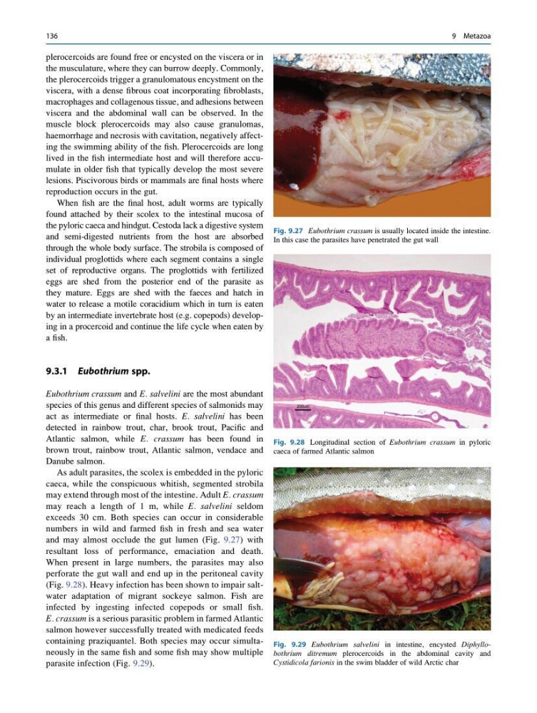
and may almost occlude the gut lumen (Fig. 9.27) with
resultant loss of performance, emaciation and death.
When present in large numbers, the parasites may also
perforate the gut wall and end up in the peritoneal cavity
(Fig. 9.28). Heavy infection has been shown to impair saltwater adaptation of migrant sockeye salmon. Fish are
infected by ingesting infected copepods or small fish.
E. crassum is a serious parasitic problem in farmed Atlantic
salmon however successfully treated with medicated feeds
containing praziquantel. Both species may occur simultaneously in the same fish and some fish may show multiple
parasite infection (Fig. 9.29).
Fig. 9.27 Eubothrium crassum is usually located inside the intestine.
In this case the parasites have penetrated the gut wall
Fig. 9.28 Longitudinal section of Eubothrium crassum in pyloric
caeca of farmed Atlantic salmon
Fig. 9.29 Eubothrium salvelini in intestine, encysted Diphyllobothrium ditremum plerocercoids in the abdominal cavity and
Cystidicola farionis in the swim bladder of wild Arctic char
136
9 Metazoa
the musculature, where they can burrow deeply. Commonly,
the plerocercoids trigger a granulomatous encystment on the
viscera, with a dense fibrous coat incorporating fibroblasts,
macrophages and collagenous tissue, and adhesions between
viscera and the abdominal wall can be observed. In the
muscle block plerocercoids may also cause granulomas,
haemorrhage and necrosis with cavitation, negatively affecting the swimming ability of the fish. Plerocercoids are long
lived in the fish intermediate host and will therefore accumulate in older fish that typically develop the most severe
lesions. Piscivorous birds or mammals are final hosts where
reproduction occurs in the gut.
When fish are the final host, adult worms are typically
found attached by their scolex to the intestinal mucosa of
the pyloric caeca and hindgut. Cestoda lack a digestive system
and semi-digested nutrients from the host are absorbed
through the whole body surface. The strobila is composed of
individual proglottids where each segment contains a single
set of reproductive organs. The proglottids with fertilized
eggs are shed from the posterior end of the parasite as
they mature. Eggs are shed with the faeces and hatch in
water to release a motile coracidium which in turn is eaten
by an intermediate invertebrate host (e.g. copepods) developing in a procercoid and continue the life cycle when eaten by
a fish.
9.3.1 Eubothrium spp.
Eubothrium crassum and E. salvelini are the most abundant
species of this genus and different species of salmonids may
act as intermediate or final hosts. E. salvelini has been
detected in rainbow trout, char, brook trout, Pacific and
Atlantic salmon, while E. crassum has been found in
brown trout, rainbow trout, Atlantic salmon, vendace and
Danube salmon.
As adult parasites, the scolex is embedded in the pyloric
caeca, while the conspicuous whitish, segmented strobila
may extend through most of the intestine. Adult E. crassum
may reach a length of 1 m, while E. salvelini seldom
exceeds 30 cm. Both species can occur in considerable
numbers in wild and farmed fish in fresh and sea water
and may almost occlude the gut lumen (Fig. 9.27) with
resultant loss of performance, emaciation and death.
When present in large numbers, the parasites may also
perforate the gut wall and end up in the peritoneal cavity
(Fig. 9.28). Heavy infection has been shown to impair saltwater adaptation of migrant sockeye salmon. Fish are
infected by ingesting infected copepods or small fish.
E. crassum is a serious parasitic problem in farmed Atlantic
salmon however successfully treated with medicated feeds
containing praziquantel. Both species may occur simultaneously in the same fish and some fish may show multiple
parasite infection (Fig. 9.29).
Fig. 9.27 Eubothrium crassum is usually located inside the intestine.
In this case the parasites have penetrated the gut wall
Fig. 9.28 Longitudinal section of Eubothrium crassum in pyloric
caeca of farmed Atlantic salmon
Fig. 9.29 Eubothrium salvelini in intestine, encysted Diphyllobothrium ditremum plerocercoids in the abdominal cavity and
Cystidicola farionis in the swim bladder of wild Arctic char
136
9 Metazoa