

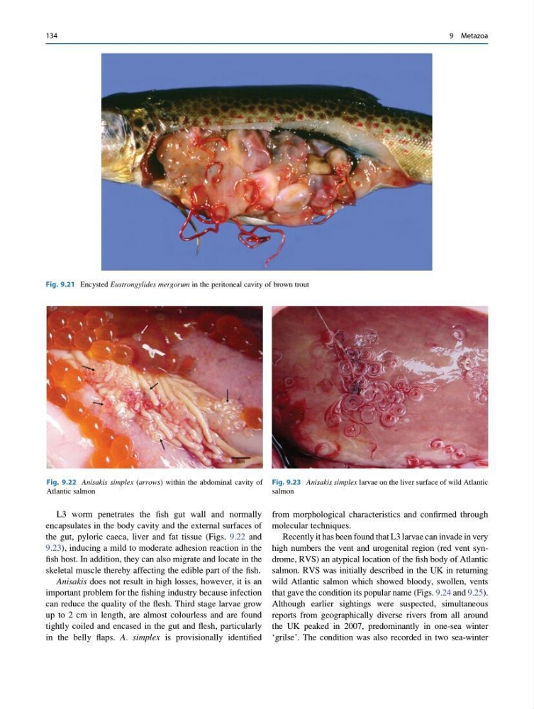
L3 worm penetrates the fish gut wall and normally
encapsulates in the body cavity and the external surfaces of
the gut, pyloric caeca, liver and fat tissue (Figs. 9.22 and
9.23), inducing a mild to moderate adhesion reaction in the
fish host. In addition, they can also migrate and locate in the
skeletal muscle thereby affecting the edible part of the fish.
Anisakis does not result in high losses, however, it is an
important problem for the fishing industry because infection
can reduce the quality of the flesh. Third stage larvae grow
up to 2 cm in length, are almost colourless and are found
tightly coiled and encased in the gut and flesh, particularly
in the belly flaps. A. simplex is provisionally identified
from morphological characteristics and confirmed through
molecular techniques.
Recently it has been found that L3 larvae can invade in very
high numbers the vent and urogenital region (red vent syndrome, RVS) an atypical location of the fish body of Atlantic
salmon. RVS was initially described in the UK in returning
wild Atlantic salmon which showed bloody, swollen, vents
that gave the condition its popular name (Figs. 9.24 and 9.25).
Although earlier sightings were suspected, simultaneous
reports from geographically diverse rivers from all around
the UK peaked in 2007, predominantly in one-sea winter
‘grilse’. The condition was also recorded in two sea-winter
Fig. 9.21 Encysted Eustrongylides mergorum in the peritoneal cavity of brown trout
Fig. 9.22 Anisakis simplex (arrows) within the abdominal cavity of
Atlantic salmon
Fig. 9.23 Anisakis simplex larvae on the liver surface of wild Atlantic
salmon
134
9 Metazoa
encapsulates in the body cavity and the external surfaces of
the gut, pyloric caeca, liver and fat tissue (Figs. 9.22 and
9.23), inducing a mild to moderate adhesion reaction in the
fish host. In addition, they can also migrate and locate in the
skeletal muscle thereby affecting the edible part of the fish.
Anisakis does not result in high losses, however, it is an
important problem for the fishing industry because infection
can reduce the quality of the flesh. Third stage larvae grow
up to 2 cm in length, are almost colourless and are found
tightly coiled and encased in the gut and flesh, particularly
in the belly flaps. A. simplex is provisionally identified
from morphological characteristics and confirmed through
molecular techniques.
Recently it has been found that L3 larvae can invade in very
high numbers the vent and urogenital region (red vent syndrome, RVS) an atypical location of the fish body of Atlantic
salmon. RVS was initially described in the UK in returning
wild Atlantic salmon which showed bloody, swollen, vents
that gave the condition its popular name (Figs. 9.24 and 9.25).
Although earlier sightings were suspected, simultaneous
reports from geographically diverse rivers from all around
the UK peaked in 2007, predominantly in one-sea winter
‘grilse’. The condition was also recorded in two sea-winter
Fig. 9.21 Encysted Eustrongylides mergorum in the peritoneal cavity of brown trout
Fig. 9.22 Anisakis simplex (arrows) within the abdominal cavity of
Atlantic salmon
Fig. 9.23 Anisakis simplex larvae on the liver surface of wild Atlantic
salmon
134
9 Metazoa