

9.2
Nematoda
The Nematoda comprise numerous species of unsegmented
roundworms that possess a pseudocoelom and a threadlike
body that is circular in cross-section. Nematodes in fish can
be found both as larvae or adult endoparasites. Nematode
infection causes a variable but generally not extensive
pathology, although more prominent where the parasite is
migrating or degenerating (Fig. 9.18). Nematode digestive
tract is divided into mouth (buccal) capsule, oesophagus,
intestine and rectum. Histologically, a multi-layered cuticle
which can be ornamented with ridges is recorded. The hypodermis is often thinner than the somatic musculature often
with extensions called lateral chords protruding into the
pseudocoelom. The musculature is composed of dense contractile elements and pale sarcoplasm. In section the oesophagus is recognised by the radial symmetry and triradiate
lumen. The structure of the epithelial cells range from
large multinucleate cells to cuboidal or tall columnar cells
and can be been used to differentiate groups.
9.2.1 Philonema oncorhynchi
Philonema oncorhynchi (Philometridae) is noted in the visceral
cavity of several fish species including rainbow trout, Arctic
char, chum salmon and sockeye salmon in freshwater from
various regions including Canada, Iceland, Japan and parts of
Europe. P. sibirica is also described from European salmonids.
Larva, sub-adult and adult worms can be present but are not
usually fatal.
Juvenile fish acquire the nematode in freshwater by feeding on copepods, i.e. Cyclops spp. that act as the intermediate
host. Larvae are released from gravid females present in the
body cavity of affected hosts at spawning, and infect a copepod where they moult twice in their haemocoel into third
stage larvae. The cycle is completed when the copepods are
eaten by the final host. The orientation of seaward-migrating
sockeye smolts is thought to be influenced by the presence of
this parasite, which may have implications for smolt survival.
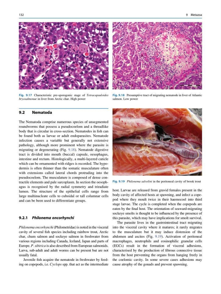
The parasite lives in the gastrointestinal tract migrating
into the visceral cavity where it matures; it rarely migrates
to the musculature but it may induce distension of the
abdomen and ascites (Fig. 9.19). Activation of peritoneal
macrophages, neutrophils and eosinophilic granular cells
(EGCs) result in the formation of visceral adhesions,
characterised by the production of fibrous connective tissue
from the host preventing the organs from hanging freely in
the coelomic cavity. In some severe cases adhesions may
cause atrophy of the gonads and prevent spawning.
Fig. 9.18 Presumptive tract of migrating nematode in liver of Atlantic
salmon. Low power
Fig. 9.19 Philonema salvelini in the peritoneal cavity of brook trout
Fig. 9.17 Characteristic pre-sporogonic stage of Tetracapsuloides
bryosalmonae in liver from Arctic char. High power
132
9 Metazoa
Nematoda
The Nematoda comprise numerous species of unsegmented
roundworms that possess a pseudocoelom and a threadlike
body that is circular in cross-section. Nematodes in fish can
be found both as larvae or adult endoparasites. Nematode
infection causes a variable but generally not extensive
pathology, although more prominent where the parasite is
migrating or degenerating (Fig. 9.18). Nematode digestive
tract is divided into mouth (buccal) capsule, oesophagus,
intestine and rectum. Histologically, a multi-layered cuticle
which can be ornamented with ridges is recorded. The hypodermis is often thinner than the somatic musculature often
with extensions called lateral chords protruding into the
pseudocoelom. The musculature is composed of dense contractile elements and pale sarcoplasm. In section the oesophagus is recognised by the radial symmetry and triradiate
lumen. The structure of the epithelial cells range from
large multinucleate cells to cuboidal or tall columnar cells
and can be been used to differentiate groups.
9.2.1 Philonema oncorhynchi
Philonema oncorhynchi (Philometridae) is noted in the visceral
cavity of several fish species including rainbow trout, Arctic
char, chum salmon and sockeye salmon in freshwater from
various regions including Canada, Iceland, Japan and parts of
Europe. P. sibirica is also described from European salmonids.
Larva, sub-adult and adult worms can be present but are not
usually fatal.
Juvenile fish acquire the nematode in freshwater by feeding on copepods, i.e. Cyclops spp. that act as the intermediate
host. Larvae are released from gravid females present in the
body cavity of affected hosts at spawning, and infect a copepod where they moult twice in their haemocoel into third
stage larvae. The cycle is completed when the copepods are
eaten by the final host. The orientation of seaward-migrating
sockeye smolts is thought to be influenced by the presence of
this parasite, which may have implications for smolt survival.
The parasite lives in the gastrointestinal tract migrating
into the visceral cavity where it matures; it rarely migrates
to the musculature but it may induce distension of the
abdomen and ascites (Fig. 9.19). Activation of peritoneal
macrophages, neutrophils and eosinophilic granular cells
(EGCs) result in the formation of visceral adhesions,
characterised by the production of fibrous connective tissue
from the host preventing the organs from hanging freely in
the coelomic cavity. In some severe cases adhesions may
cause atrophy of the gonads and prevent spawning.
Fig. 9.18 Presumptive tract of migrating nematode in liver of Atlantic
salmon. Low power
Fig. 9.19 Philonema salvelini in the peritoneal cavity of brook trout
Fig. 9.17 Characteristic pre-sporogonic stage of Tetracapsuloides
bryosalmonae in liver from Arctic char. High power
132
9 Metazoa