

Throughout the affected tissue characteristic PKX cells
can be found, particularly in the endothelium of the portal
vessels. These cells are large, eosinophilic to pale orange
(often multinucleated), PAS-positive cells with a granular
cytoplasm and typically surrounded by a clear halo. PKX
cells may be seen in several other organs including the gills,
spleen, liver and heart. Developmental stages (sporoblasts)
of the parasite may also be recorded in the walls and lumen
of the kidney tubules.
The diagnosis of PKD is based on clinical signs and the
demonstration of the characteristic extrasporogonic stages in
wet preparations, stained imprints or in histological sections.
In addition, a specific PCR assessment can be used as a
confirming test. Sphaerospora truttae is important from the
viewpoint of differential diagnostics.
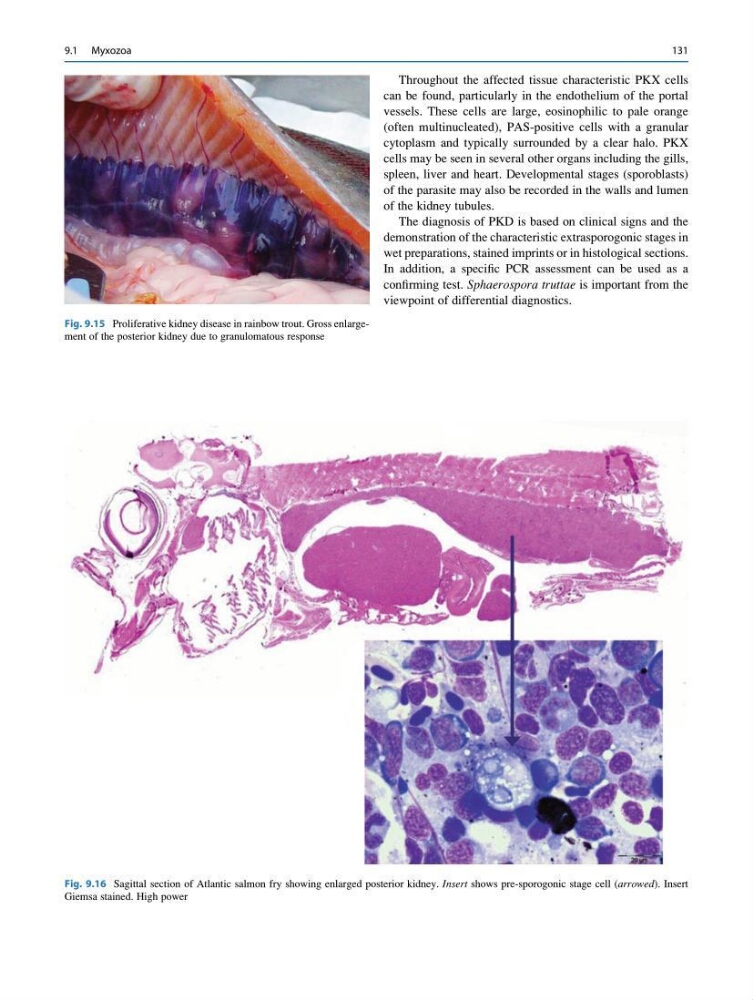
Fig. 9.15 Proliferative kidney disease in rainbow trout. Gross enlargement of the posterior kidney due to granulomatous response
Fig. 9.16 Sagittal section of Atlantic salmon fry showing enlarged posterior kidney. Insert shows pre-sporogonic stage cell (arrowed). Insert
Giemsa stained. High power
9.1 Myxozoa
131
can be found, particularly in the endothelium of the portal
vessels. These cells are large, eosinophilic to pale orange
(often multinucleated), PAS-positive cells with a granular
cytoplasm and typically surrounded by a clear halo. PKX
cells may be seen in several other organs including the gills,
spleen, liver and heart. Developmental stages (sporoblasts)
of the parasite may also be recorded in the walls and lumen
of the kidney tubules.
The diagnosis of PKD is based on clinical signs and the
demonstration of the characteristic extrasporogonic stages in
wet preparations, stained imprints or in histological sections.
In addition, a specific PCR assessment can be used as a
confirming test. Sphaerospora truttae is important from the
viewpoint of differential diagnostics.
Fig. 9.15 Proliferative kidney disease in rainbow trout. Gross enlargement of the posterior kidney due to granulomatous response
Fig. 9.16 Sagittal section of Atlantic salmon fry showing enlarged posterior kidney. Insert shows pre-sporogonic stage cell (arrowed). Insert
Giemsa stained. High power
9.1 Myxozoa
131