

in the northwest coast of the USA (Puget Sound, Washington
region). Elevated pre-spawning mortality in some of these
species has been associated to infection with P. minibicornis.
Clinical signs and pathological changes are unspecific and
may include dark and lethargic fish with hypertrophied kidney
(Fig. 9.13). Histologically, trophozoites and developing
spores occur in the lumen and epithelium. A PCR assay
has identified the myxospore in the freshwater polychaete,
Manayunkia speciosa.
Parvicapsula kabatai has been described from renal tubules
of pink salmon in British Columbia, Canada. The shape and
size of the spores are similar to those of P. pseudobranchicola,
but distinctly different from P. minibicornis. The significance
of this parasite is currently unknown.
9.1.8 Sphaerospora truttae
Sphaerospora truttae was originally described from brown
trout and grayling in Germany, but subsequently reported
affecting and Atlantic salmon parr in Scotland. Brown trout
are also proven susceptible. The gills have been identified
as the predominant point of entry, which is followed by
penetration of the vascular epithelia and thereafter, proliferation in the blood before exiting the vascular system
through capillary walls. Subsequently, the kidney, as well
as the spleen and the liver are infected. Parasites occur in
the tubular lumen and sporogony takes place inside the
renal tubules (Fig. 9.14). Histology can be used for presumptive identification but for early myxosporean stages
and parasite specific identity, a DNA-based approach is
appropriate.
9.1.9 Tetracapsuloides bryosalmonae
Tetracapsuloides bryosalmonae causes the condition proliferative kidney disease (PKD) which is a significant seasonal
disease of young salmonids. PKD occurs both in farmed and
wild fish and is associated with decline in wild populations in
many countries. The endoparasitic myxozoan uses freshwater
bryozoans as primary hosts. Environmental changes may play
a role in the increased significance of PKD in wild populations.
Bryozoans and T. bryosalmonae stages in bryozoans undergo
temperature and nutrient-driven proliferation and above 15
C
are required for development of clinical disease. Infective
spores enter the fish through the skin and gill epithelium.
Clinical signs include a dark body, bilateral exophthalmia, pale gills, and distended abdomen with pale
visceral organs. Swelling due to extensive accumulation of
ascites is recorded within the abdomen. The kidney, particularly the caudal region, is markedly swollen due to diffuse
oedema (Fig. 9.15) and to a lesser extent the spleen.
Histozoic and extrasporogonic stages proliferate causing
focal or multifocal granulomatous inflammation and renal
interstitial tissue is replaced by mild haematopoietic hyperplasia during the early stages of infection, and followed by
further granulomatous tissue with associated macrophages
and mononuclear cells. Lymphoid cells and macrophages
are frequently seen adherent to the PKX cells (Fig. 9.16)
and hepatic lesions frequently include multinucleated giant
cells scattered throughout (Fig. 9.17). There may also be
extensive haemorrhage in the acute stages of the disease.
The number of nephrons and melanomacrophage centres are
severely reduced and an extensive chronic fibrosis occurs in
the final stages.
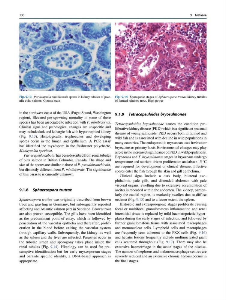
Fig. 9.13 Parvicapsula minibicornis spores in kidney tubules of juvenile coho salmon. Giemsa stain
Fig. 9.14 Sporogonic stages of Sphaerospora truttae kidney tubules
of farmed rainbow trout. High power
130
9 Metazoa
region). Elevated pre-spawning mortality in some of these
species has been associated to infection with P. minibicornis.
Clinical signs and pathological changes are unspecific and
may include dark and lethargic fish with hypertrophied kidney
(Fig. 9.13). Histologically, trophozoites and developing
spores occur in the lumen and epithelium. A PCR assay
has identified the myxospore in the freshwater polychaete,
Manayunkia speciosa.
Parvicapsula kabatai has been described from renal tubules
of pink salmon in British Columbia, Canada. The shape and
size of the spores are similar to those of P. pseudobranchicola,
but distinctly different from P. minibicornis. The significance
of this parasite is currently unknown.
9.1.8 Sphaerospora truttae
Sphaerospora truttae was originally described from brown
trout and grayling in Germany, but subsequently reported
affecting and Atlantic salmon parr in Scotland. Brown trout
are also proven susceptible. The gills have been identified
as the predominant point of entry, which is followed by
penetration of the vascular epithelia and thereafter, proliferation in the blood before exiting the vascular system
through capillary walls. Subsequently, the kidney, as well
as the spleen and the liver are infected. Parasites occur in
the tubular lumen and sporogony takes place inside the
renal tubules (Fig. 9.14). Histology can be used for presumptive identification but for early myxosporean stages
and parasite specific identity, a DNA-based approach is
appropriate.
9.1.9 Tetracapsuloides bryosalmonae
Tetracapsuloides bryosalmonae causes the condition proliferative kidney disease (PKD) which is a significant seasonal
disease of young salmonids. PKD occurs both in farmed and
wild fish and is associated with decline in wild populations in
many countries. The endoparasitic myxozoan uses freshwater
bryozoans as primary hosts. Environmental changes may play
a role in the increased significance of PKD in wild populations.
Bryozoans and T. bryosalmonae stages in bryozoans undergo
temperature and nutrient-driven proliferation and above 15
C
are required for development of clinical disease. Infective
spores enter the fish through the skin and gill epithelium.
Clinical signs include a dark body, bilateral exophthalmia, pale gills, and distended abdomen with pale
visceral organs. Swelling due to extensive accumulation of
ascites is recorded within the abdomen. The kidney, particularly the caudal region, is markedly swollen due to diffuse
oedema (Fig. 9.15) and to a lesser extent the spleen.
Histozoic and extrasporogonic stages proliferate causing
focal or multifocal granulomatous inflammation and renal
interstitial tissue is replaced by mild haematopoietic hyperplasia during the early stages of infection, and followed by
further granulomatous tissue with associated macrophages
and mononuclear cells. Lymphoid cells and macrophages
are frequently seen adherent to the PKX cells (Fig. 9.16)
and hepatic lesions frequently include multinucleated giant
cells scattered throughout (Fig. 9.17). There may also be
extensive haemorrhage in the acute stages of the disease.
The number of nephrons and melanomacrophage centres are
severely reduced and an extensive chronic fibrosis occurs in
the final stages.
Fig. 9.13 Parvicapsula minibicornis spores in kidney tubules of juvenile coho salmon. Giemsa stain
Fig. 9.14 Sporogonic stages of Sphaerospora truttae kidney tubules
of farmed rainbow trout. High power
130
9 Metazoa